Science Photography
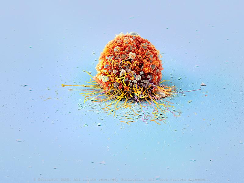
Micronaut creates award-winning micrographs of invisibly small organisms and structures. Often the corresponding samples are collected from every corner of the Earth, or are cultured and carefully put on stage under laboratory conditions (in vitro), similar to the picture of an invasively growing cancer cell (Best Scientific Image, Fascination Research; 1st prize).
This series of works contains images that were made to visualize key aspects and newest results achieved by modern scientific research. Most images have been produced in close collaboration with the corresponding scientists, and research area experts.
«These images are really outstanding. They raise all sorts of research questions I had never even considered before.»
Rob R. Dunn, PhD
Prof. of Applied Ecology and Book author
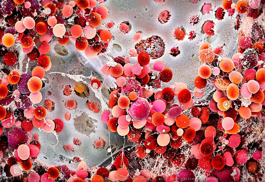
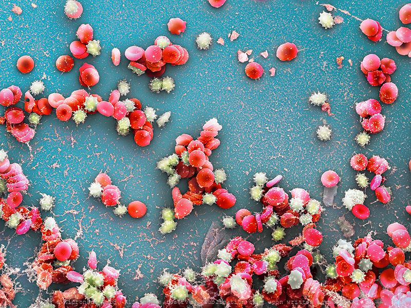
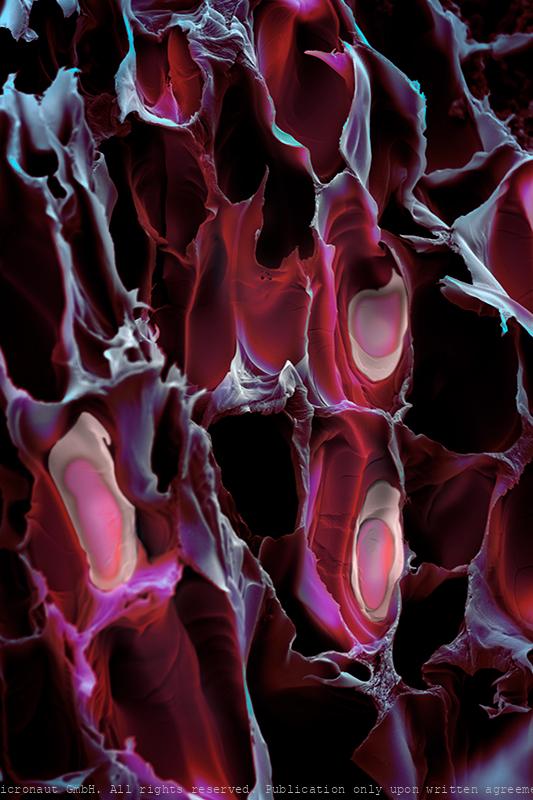
Blood- and bone marrow stem cells (Homo sapiens) - Knochenmarkas
A stem cell or bone marrow transplant replaces damaged blood cells with healthy ones. It can be used to treat diseases such as leukaemia and lymphoma, which affect the blood cells of the patient. After removing damaged blood cells, healthy cells can be derived from blood- or bone-marrow (a spongy tissue found in the centre of some bones) stem cells. Stem cells can turn into different types of blood cells, including: - red blood cells – which carry oxygen around the body - white blood cells – which help fight infection - platelets – which help stop bleeding. By Martin Oeggerli, supported by SRK, Andi Buser and Andi Holbro, and Pathology Univ. Hosp. Basel, and Bio-EM Lab, Univ. Basel.
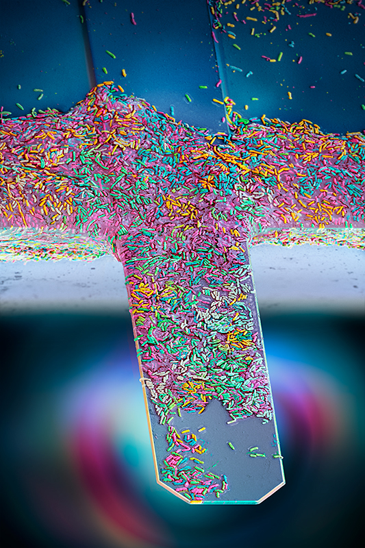
Nanomotion sensor - detecting the vibration of living bacteria i
Antibiotic Resistance Chip Loaded with Bacteria. Hand-colored SEM scan by Martin Oeggerli / Micronaut, 2021. Antibiotic resistance is currently one of biggest threats to global health. By offering a diagnostic device, Resistell proposes an alternative to culture based antibiogram, the current gold standard in antibiotic susceptibility testing. The rapid AST method is based on the detection of movement caused by living bacterial cells. Because the test is growth independent rapid AST, we reduce the time taken to get a result from days to minutes. Resistell provides information on which antibiotic should be used to treat the patient, and the concentration at which it should be administered. This rapid AST designed and developed by Resistell saves lives by finding the right antibiotic on time and reduces spreading of antibiotic resistance!
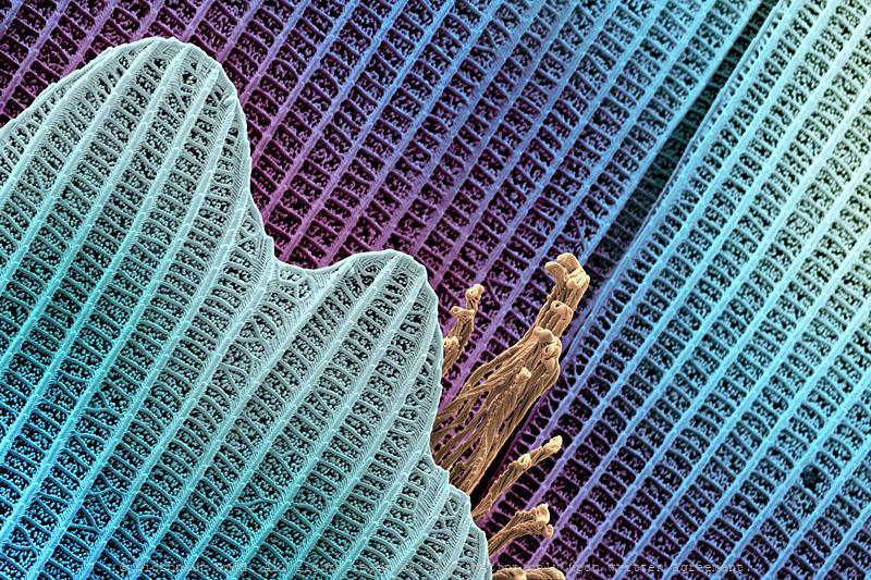
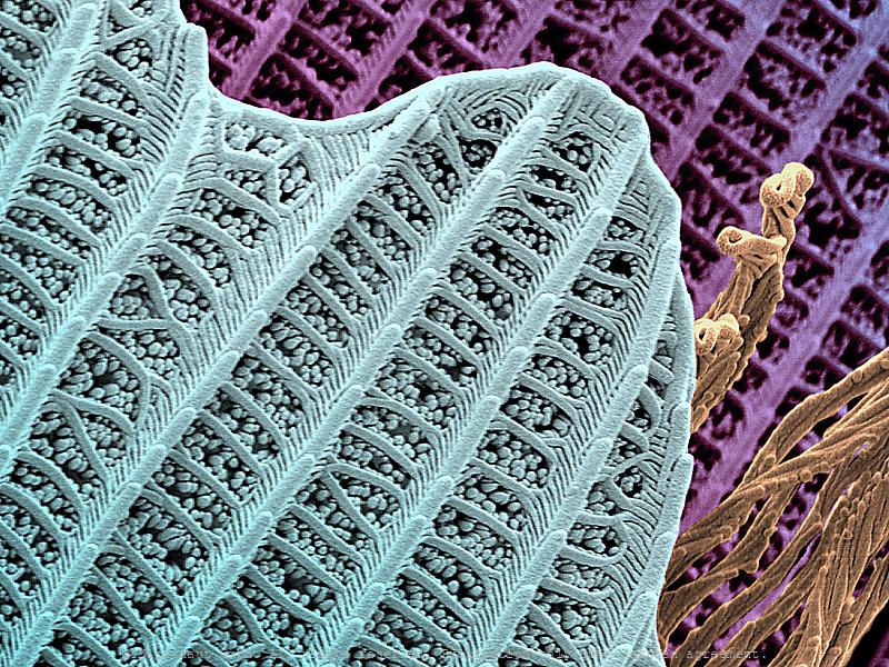
Pheaodactylum tricornutum
Pheaodactylum tricornutum
Synthetic mini hearts beat like the real ting!
Advancements in the development of patented synthetic human-like hearts allow researchers to study the development of the human heart and related disease on highly accurate models. This facilitates the development of innovative therapies and new pharmaceutical drugs to treat heart-related problems such as cardiovascular and metabolic diseases. Hand-colored scanning electron micrograph (SEM) by Martin Oeggerli in collaboration with Makoto Hara and Lucas Goguen of the Novartis Institute for BioMedical Research (NIBR). Annually an estimated 21 million deaths are related to disorders of the heart and blood vessels, and that number is growing. By creating synthetic organoids that beat like the real thing, scientists are unravelling the intricacies of the heart. Similar in size and development to fetal human hearts, these mini heart organoids are becoming increasingly complex and realistic.
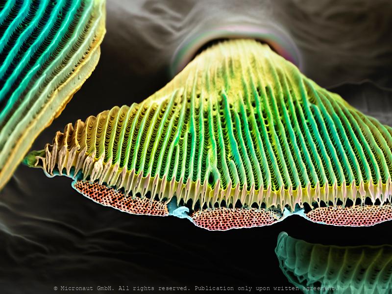
Solar Powered Future (C. staurestrum)
Image shows C. Staurestrum algae from Prof. Ben Hankamer's multidisciplinary approach to use light-driven microalgae chemistry to convert solar energy into sustainable products and ecosystem services. This breakthrough “third way” of harnessing the sun's power goes beyond electricity and heat, transforming sunlight directly into renewable fuels, food and clean water. The team’s work addresses urgent global challenges including CO₂ reduction, food security and water purification, while leveraging Australia’s natural strengths — sunlight, space and scientific talent. With solar energy capable of powering the world in just two hours, solar biotechnologies offer a scalable path to new green industries, jobs, and exports. Driven by purpose and innovation, Professor Hankamer’s research positions Australia as a global leader in sustainable solar solutions, demonstrating how science can turn energy abundance into environmental and economic resilience.
Satellite Cell (Mus musculus) - The Stem Cell that Regenerates t
2021 marks the 60th anniversary of the discovery of the "satellite cell" by Alexander Mauro by using the electron microscope and it seems an opportune moment for two researchers from the University Basel to team up with Micronaut and present a state of the art picture of a muscular stem cell. To produce this picture, mouse muscles were digested and cultured by Sedat Dilbaz and Gesa Santos in the lab of Prof. Christoph Handschin, at the Biozentrum, University Basel. Once satellite cells got activated, the sample was carefully fixed and a scanning electron microscope (SEM) was used to capture black-and-white SEM scans. After this, photorealistic colors were added in the digital darkroom by the artist. The basic unit of skeletal muscle is the myofiber: a syncytial cell packed with myofibrils, containing the sarcomeres that generate force by contraction. In vertebrates, each myofibre is controlled by many (usually hundreds) of myonuclei, which in mammals are considered post mitotic. During growth, new myonuclei are supplied by muscle satellite cells: resident stem cells located on the surface of a myofibre. In mature muscles, satellite cells become quiescent ("dormant"), but remain able to provide myoblasts for muscle hypertrophy and repair if needed. That muscle is capable of regeneration was first shown in the 1860s, but almost a century elapsed before the satellite cell was discovered. Electron microscopic studies by Alexander Mauro identified the satellite cells wedged between the plasma membrane of the muscle fibre and basement membrane and revealed that they almost completely lack cytoplasm. While nowadays, the muscle satellite cell is widely accepted as the resident stem cell of skeletal muscle, supplying myoblasts for growth, homeostasis and repair, less is known about how Satellite cells remain quiescent, or how they are activated. The understanding of the complex cellular and molecular interactions of satellite cells with their dynamic microenvironment rem
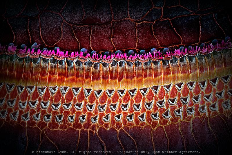
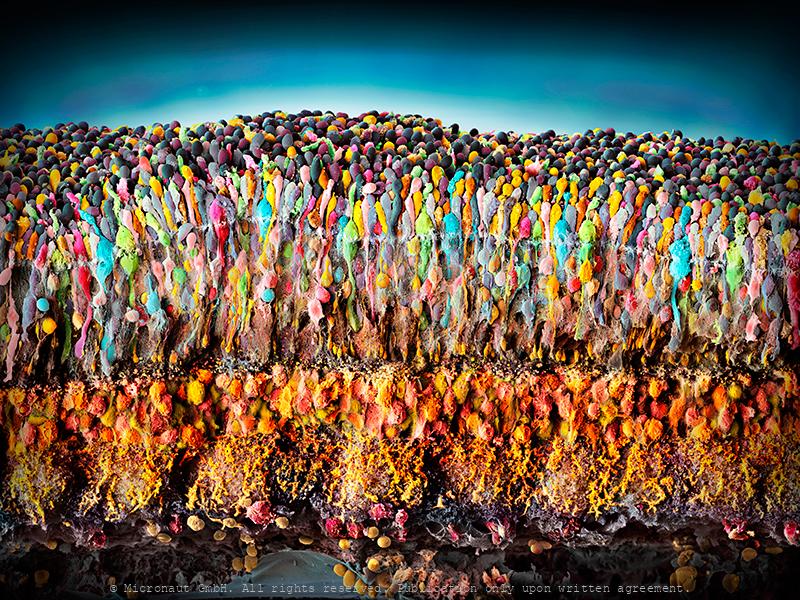
Porcine retina (Sus domesticus)
Porcine Retina (Sus domesticus) With far more than 100 million nerve cells, the retina is our first stage of the visual system and window to the outside world. The picture highlights the neuronal layers of a porcine retina. Seungkuk Ahn and his colleagues have electromagnetically guided viruses to specifically transduce each of the retina layers. The process inside the retina includes detection of light impulses (photons) by different light receptors, in general called rods (120 million cells) and cones (6 million cells), and the fast and continuous translation, filtration and post-procession into electrical signals (or nerve impulses). These signals are then passed through the optical nerve’s 1.5 million fibres to the visual centre of the brain and reinterpreted into a cohesive image.The flexibility and economy with which retinal cells work together remains beyond our powers of imagination: the eye reports numerous signals to the brain at once, including separate detection of light (light on) and dark (light off), general patterns and finest details, movements and different hues (including: red, green and blue). This literally makes the eye a camera with 10 to 15 different films and despite it appears so naturally to us, the perception of an image is the result of a most complex interaction process involving millions of cells and the electrical signals they produce and pass on to the brain.
Hearing Sensors (Mus musculus)
Hearing Sensors This image shows the sensory epithelium of the inner ear (Organ of Corti), with the apical protruding hair bundle of hair cells. Scanning-electron-microscopic (SEM) image of a 22-week-old wild-type C57BL/6J mouse, carefully colored to harmonize with the natural shadows of the image and to highlight the invisibly small structures that are responsible for hearing in mammals. The organ of Corti was called after the Italian scientist (Alfonso Corti), who first described it. Its three rows of highly ordered hair cells, located inside the cochlea, are unique among other organs of the body and generate nerve impulses in response to sound vibrations. By Maurizio Cortada and Martin Oeggerli. Supported by Pathology, Univ. Hosp. Basel, and Bio-EM Lab, Univ. Basel.
Der Zauberpilz - The Psycodelic Mushroom (Claviceps purpurea)
Lysergic acid diethylamide is a synthetic lysergamide that can be obtained as a derivative of naturally occurring ergot alkaloids. LSD is one of the most powerful hallucinogens known and belongs to a subgroup of psychedelics that affect the body's serotonin system. In mid-April 1943, the Swiss chemist Albert Hofmann accidentally discovered the psychoactive substance LSD. The drug has since been banned, but in the right dosage and with therapeutic support, LSD can also be used as a helpful medication against depression. Thinking, feeling and perception are massively influenced by the psychoactive substance, the experience of space and time changes, sensory illusions, colors, image distortions or delusions may occur. -- Lysergsäurediethylamid ist ein synthetisches Lysergamid, das als Derivat natürlich vorkommender Mutterkornalkaloiden erhalten werden kann. LSD ist eines der stärksten bekannten Halluzinogene und gehört zu deren Teilgruppe der Psychedelika, welche auf das Serotonin-System des Körpers wirken. Mitte April 1943 entdeckte der Schweizer Chemiker Albert Hofmann per Zufall die psychoaktive Substanz LSD. Zwischenzeitlich wurde die Droge verboten, doch in richtiger Dosierung und in therapeutischer Begleitung kann LSD auch als hilfreiches Medikament gegen Depressionen eingesetzt werden. Denken, Fühlen und die Wahrnehmung werden durch die psychoaktive Substanz massiv beeinflusst, das Erleben von Raum und Zeit verändert sich, es kann zu Sinnestäuschungen, bunten Farben, Bildverzerrungen und Wahnvorstellungen kommen.
human Microbiome of a KISS
The Human Microbiome of a Kiss. Artwork created by Martin Oeggerli (Micronaut) and dedicated to his 8-years old son Nelson. This image was originally produced for the National Geographic feature article 'How trillions of microbes affect every stage of our life-from birth to old age'. Soon after its public release, our daily life became heavily overshadowed by the world's coronavirus (COVID-19) pandemic - thereby instantly confirming the articles visionary title. Meanwhile (at end of April 2020), the crisis has not only severely limited the artists ability to work, but also prohibits him from officially visiting his son who is living in Switzerland, just a stone's throw away from the rest of the father's German-based patch-work family. This picture is based on scanning-electron-microscopy technology, and all colors are manually added by the artist in post-production, to visualize the immense diversity of the microbial community transmitted through a kiss. Whenever we kiss someone on the lips or cheeks invisibly small microbes are exchanged from one person to the other one! The variety of microbes that are part of our microbiome is mind-blowing, and the composition of the invisibly small communities can be the cause of many factors, including the human genetic makeup, diet, age, surroundings, and sexual behavior. Among humans, approximately 90% of cultures have some type of kissing. Usually it is platonic, such as a parent kissing a child. However, in 77 of 168 (46%) of all cultures, it can go as far as intimate (French-) kissing. Recently, a scientific study has revealed that on average 80 million bacteria are transferred to the partner during a kiss of 10 s. Most partners share a more similar oral microbiome compared to unrelated individuals and/or e.g. their kids. But some of the collective bacteria among partners are only transiently present, while others have found a true niche and survive permanently, allowing long-term colonization.With regard to kissing, th
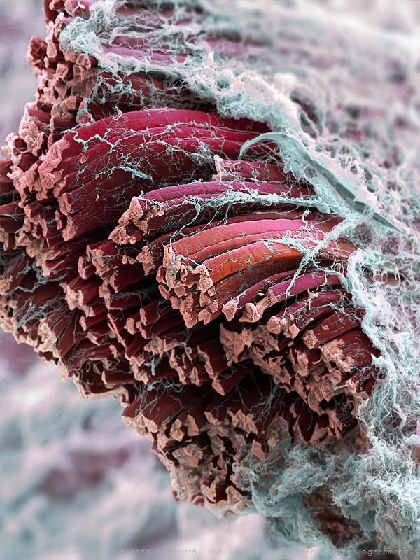
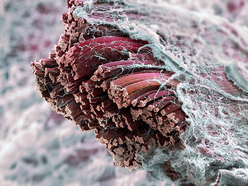
Skeletal muscle fibers (Mammalia)
Mouse skeletal muscle fibers – The image was prepared in collaboration with Sarina Meinen and colleagues from Markus Rüegg’s lab at the Biozentrum Basel, and recently published on the cover of The EMBO Journal. Connective tissue and extracellular components (white) surround the muscle, which consists of individual muscle fibres of different types (red). Sponsor Image of the month
Bloodclot / Blutgerinnsel
Red blood cells are the most common type of blood cell and are the vertebrate body's principal means of delivering oxygen from the lungs or gills to body tissues via the blood. Folllowing an injury, blood clotting produces a fibrin network which closes the wound. Induced blood clotting can also lead arterial embolism and incuce cerebrovascular accidents, including strokes.
Quasi una Fantasia - The Dancing Hamster Cell (2/3)
Tracing back a hamster’s journey from the deserts of China to the research labs of Harvard in the mid-50s and on to becoming the golden standard for the bio-manufacturing industry and eventually - a work of art. Since they were first isolated in the late 1950s, Chinese hamster ovary (CHO) cells travel on an amazing journey. Because they are so easy to grow much was already known about their genetics and growth characteristics, CHO cells moved into the spotlight of the growing bio-pharmaceutical industry, which is currently worth more than $ 125 billion per year in sales at high profit margins. The step that catapulted CHO cells into prominence was the FDA approval for bio-therapeutic protein production - combined with an ability to grow in suspension, which greatly facilitates cell culturing at large scale. Meanwhile, CHO have become the golden standard for the production of therapeutic proteins, including humanized antibodies, and continue an unbelievable voyage all around the world. This hand-colored scanning-electron-micrograph (SEM) shows a chinese hamster (Cricetulus griseus) ovary cell on a phonograph record of Ludwig van Beethoven’s Moonlight Sonata (Sonata No 14 in C). The phonograph record was the primary medium used to store and reproduce music from the late 1880s. Records were later mainly made of Polyvinyl and retained the largest market share for about 100 years. Even after the introduction of the compact disc, and other new digital media formats, the Vinyls continue to be used and listened by DJs and a growing niche market of audiophiles… and they even serve as a perfect stage to present the worlds most famous cell - as work of art.
Quasi una Fantasia - The Dancing Hamster Cell (3/3)
Tracing back a hamster’s journey from the deserts of China to the research labs of Harvard in the mid-50s and on to becoming the golden standard for the bio-manufacturing industry and eventually - a work of art. Since they were first isolated in the late 1950s, Chinese hamster ovary (CHO) cells travel on an amazing journey. Because they are so easy to grow much was already known about their genetics and growth characteristics, CHO cells moved into the spotlight of the growing bio-pharmaceutical industry, which is currently worth more than $ 125 billion per year in sales at high profit margins. The step that catapulted CHO cells into prominence was the FDA approval for bio-therapeutic protein production - combined with an ability to grow in suspension, which greatly facilitates cell culturing at large scale. Meanwhile, CHO have become the golden standard for the production of therapeutic proteins, including humanized antibodies, and continue an unbelievable voyage all around the world. This hand-colored scanning-electron-micrograph (SEM) shows a chinese hamster (Cricetulus griseus) ovary cell on a phonograph record of Ludwig van Beethoven’s Moonlight Sonata (Sonata No 14 in C). The phonograph record was the primary medium used to store and reproduce music from the late 1880s. Records were later mainly made of Polyvinyl and retained the largest market share for about 100 years. Even after the introduction of the compact disc, and other new digital media formats, the Vinyls continue to be used and listened by DJs and a growing niche market of audiophiles… and they even serve as a perfect stage to present the worlds most famous cell - as work of art.
Quasi una Fantasia - The Dancing Hamster Cell (1/3)
Tracing back a hamster’s journey from the deserts of China to the research labs of Harvard in the mid-50s and on to becoming the golden standard for the bio-manufacturing industry and eventually - a work of art. Since they were first isolated in the late 1950s, Chinese hamster ovary (CHO) cells travel on an amazing journey. Because they are so easy to grow much was already known about their genetics and growth characteristics, CHO cells moved into the spotlight of the growing bio-pharmaceutical industry, which is currently worth more than $ 125 billion per year in sales at high profit margins. The step that catapulted CHO cells into prominence was the FDA approval for bio-therapeutic protein production - combined with an ability to grow in suspension, which greatly facilitates cell culturing at large scale. Meanwhile, CHO have become the golden standard for the production of therapeutic proteins, including humanized antibodies, and continue an unbelievable voyage all around the world. This hand-colored scanning-electron-micrograph (SEM) shows a chinese hamster (Cricetulus griseus) ovary cell on a phonograph record of Ludwig van Beethoven’s Moonlight Sonata (Sonata No 14 in C). The phonograph record was the primary medium used to store and reproduce music from the late 1880s. Records were later mainly made of Polyvinyl and retained the largest market share for about 100 years. Even after the introduction of the compact disc, and other new digital media formats, the Vinyls continue to be used and listened by DJs and a growing niche market of audiophiles… and they even serve as a perfect stage to present the worlds most famous cell - as work of art.
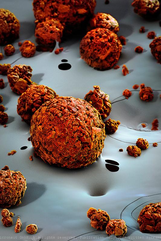
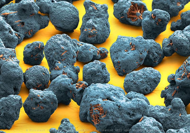
Capsugel boulders
Capsugel - Lonza - Pharmaceutical
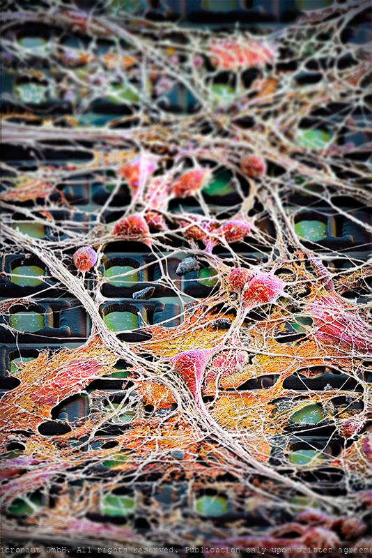
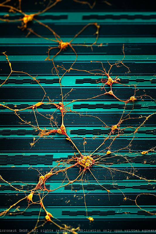
Neuronal Highways - Brain Cells on a Chip
Preparations of electro-active cells, predominantly brain cells, are placed directly atop fully processed microelectronics chips carrying thousands of electrodes. The chips are used to address fundamental questions in neuroscience and medicine. Artwork created by Martin Oeggerli (Micronaut). The picture is based on scanning-electron-microscopy technology, and all colors are manually added in post-production to visualize the research field of Electrophysiology and Neuroscience. We pursue an extracellular, bioelectronic approach to electrophysiology, which relies on the close juxtaposition of electrogenic cells (cells that produce electrical activity) and tissues with state-of-the-art integrated electronic systems. The cell preparations (dissociated cells, tissue slices) are placed directly atop fully processed microelectronics chips carrying thousands of electrodes and featuring CMOS circuitry. The bio-electronic interface consists of noble-metal electrodes. Prospective fields of applications of our technology include, besides fundamental neuroscience research, pharmascreening, the investigation of mechanisms of neurodegenerative diseases, or the development of aural and visual prostheses. Electrophysiology is the study of the electrical properties of biological cells and tissues. It involves measurements of voltage changes or electric currents on a wide variety of scales from single ion channels to whole organs like the heart. In neuroscience, it includes measurements of the electrical activity of neurons and, particularly, action potential activity. Neuroscience is the scientific study of molecular, cellular, developmental, structural, functional, evolutionary, computational, and medical aspects of the nervous system. It is an interdisciplinary science that includes aspects of biology, chemistry, computer science, engineering, mathematics and medicine.

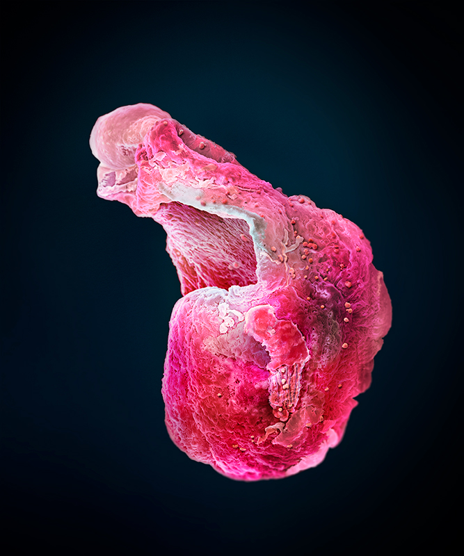
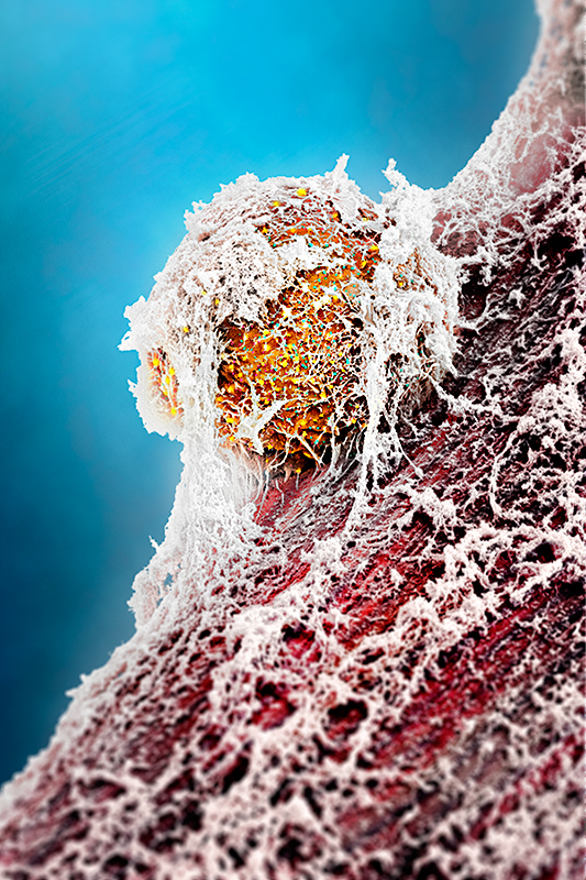
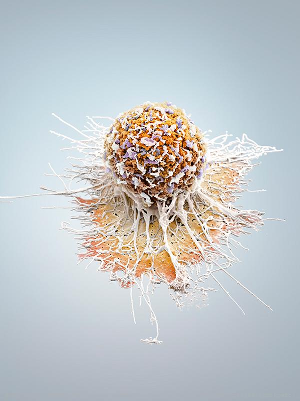
Invasively Growing Cancer Cell
Invasively growing human cancer cell, in vitro. Cancer cells develop the ability to move themselves, sooner or later. The picture shows an invasively growing tumor cell, which is pulling itself around with the help of its appendages. In comparison to a healthy cell its extensions are smaller and because of the quicker assembly and disassembly of the appendixes, it is able to move faster. -- Die Transformation einer gesunden Zelle in eine Tumorzelle ist ein komplexer Vorgang und bedingt eine ganze Reihe verschiedener genetischer Veränderungen. Tumorzellen entwickeln dabei früher oder später die Fähigkeit sich fortzubewegen und Metastasen zu bilden. Das Bild zeigt eine invasive Prostatakarzinomzelle, welche häufig Metastasen im Knochenmark bilden. Mit Hilfe der Fortsätze zieht sich die Zelle über die Unterlage. Im Vergleich zu einer gesunden Zelle bildet diese invasive Tumorzelle deutlich kleinere Fortsätze aus und ist dank schnellerem Auf- und Abbau (der Fortsätze) in der Lage, sich wesentlich schneller fortzubewegen.
Microscopic DNA-injection machine
This hand-colored scanning-electron-micrograph (SEM) shows a self-reconfiguring metamorphic nanoinjector for DNA injection into a zygote. The ability to transfer a gene or DNA sequence from one animal into the genome of another plays a critical role in a wide range of medical research—including cancer, Alzheimer's disease, and diabetes. The traditional method of transferring genetic material in-vitro into a new cell, called "microinjection," has a serious downside: it involves using a small glass pipette to pump a solution containing DNA into the nucleus of an egg cell, which results in a 25 to 40 percent cell death rate. Now, thanks to the work of researchers Brigham Young University, there's a way to avoid cell death when introducing DNA into egg cells. The MEMS nanoinjector's lance is incredibly small and no extra fluid is used with this technique, so cells undergo much less stress compared to the traditional microinjection process. "Essentially, we use electrical forces to attract and repel DNA—allowing injections to occur with a tiny, electrically conductive lance," explained Brian Jensen, associate professor in the Department of Mechanical Engineering at Brigham Young University. "DNA is attracted to the outside of the lance using positive voltage, and then the lance is inserted into a cell."
Micro Robots
Colored SEM image showing micro robots named 'PolyMites', arranged on a waver plate. They are the next generation of MagMites (magnetic mite robots). The PolyMites are not released from silicon wafer yet. In the end the robots will have a round shape without the two bars which hold each robot in place on the waver plate.
CMA432
Rotation-Micro-Sensor - CMA 432 (Bosch)
Brain Cells on a Chip (blue)
This work shows brain cells growing on a futuristic sensor-system, designed to analyse neuronal signalling. Every human thought starts with a signal traveling from one neuron to another in the brain. Yet we know relatively little about how these connections form and grow into the highly ordered neuronal circuits, thereby connecting billions of cells in the brain of a human adult. Understanding how neural circuits are formed is one of the fundamental questions in neuroscience. To study the growth characteristics and signalling, scientists have grown neuronal cells on semiconductor chip. This artwork was created by Martin Oeggerli (Micronaut). The picture is based on scanning-electron-microscopy technology. Colors are manually added by the artist in post-production to highlight the delicate interactions between brain cells.
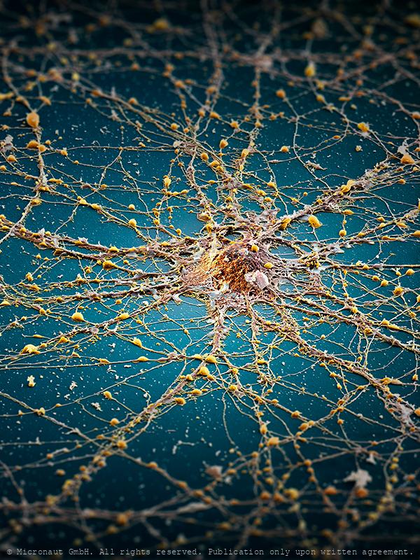
The Neuronal Megacity (Brain cell)
My concept concerning this picture was to compare a nerve cell to an island, or megacity, indicating the complex architecture of the brain. In detail, an infinite number of cellular extensions and microscopical structures unravels (capacity & complexity of the brain), whereas in the overview you can see a confusing tangle of connections that lead towards, or away, or around the center point, like the road network in a megacity. The edges of the picture are deliberately darkened and blurred, indicating there is a lot to be discovered and learned in this area of modern scientific research. Last but not least - the blue “sea” stands for Hawaii / Sophie H, to whom this picture is dedicated. Artwork created by Martin Oeggerli (Micronaut). The picture is based on scanning-electron-microscopy technology, and all colors were manually added to visualize the complex network as formed by neuronal cells in vitro.
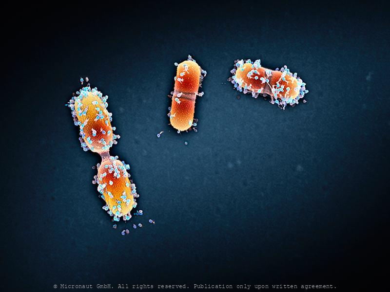
Viral infection - The Parasites of Parasites of Parasites... Nr.1
A bacteriophage is a virus that infects and replicates within a bacterium. It is composed of proteins that encapsulate a DNA or RNA genome. Bacteriophages ('phages') have relatively simple structures and their genomes encodes something between four and several hundred genes. To replicate, phages inject their genome into the cytoplasm of a bacteria. Bacteriophages are among the most common and diverse entities in the biosphere. They are widely distributed in locations populated by bacterial hosts, such as soil or the intestines of animals. One of the densest natural sources for phages and other viruses is sea water. This hand colored scanning-electron-micrograph shows bacteria which are heavily infected by bacteriophages.
Dividing Streptococcus (beta-haem; remastered version)
dividing streptococcus sp. bacteria
Skeletal muscle fibers (Mammalia)
Mouse skeletal muscle fibers – The image was prepared in collaboration with Sarina Meinen and colleagues from Markus Rüegg’s lab at the Biozentrum Basel, and recently published on the cover of The EMBO Journal. Connective tissue and extracellular components (white) surround the muscle, which consists of individual muscle fibres of different types (red). Sponsor Image of the month

Skeletal muscle fibers (Mammalia)

Acceleration-Micro-Sensor (Bosch), Nr.2
Acceleration-Micro-Sensor - CMG 262 (Bosch)
Acceleration-Micro-Sensor (Bosch), Nr.1
Acceleration-Micro-Sensor - CMG 262 (Bosch)
It's only the tongue (but I like it)
A sense of taste - taste bud & rough surface of the tongue In 1970 John Pasche designed the famous Hot Lips Logo for the rock band The Rolling Stones. 50 years later the band continues to play while this hand-colored SEM picture sheds new light on the microscopically small rough texture of the tongue. Between dozens of tiny bumps called papillae, one tip of a taste bud can be seen. The tongue is covered with moist, pink tissue called mucosa, and thousands of taste buds cover the surfaces. Each one is a collections of nerve-like cells that connect to nerves running into the brain. A stuck-out tongue can have 2'000-8'000 delicate taste buds, which are nestled beneath the surface of the tongue like sections of an orange. Only the tip of a taste bud pokes to the surface, to detect one of five main tastes (sweet, bitter, sour, salty, or savory)... Oeggerli slowly breathes life into his works by painstakingly selecting and masking different structures with color, layer upon layer utilizing his laptop touchpad. The process allows him to set focus on the mysterious microscopic landscape of an organ that we think we know best. Yet, the tongue remeins strange and unfamiliar under the SEM - almost like it was an extra-terrestrial landscape. -- Geschmacksknospe & Oberfläche der Zunge
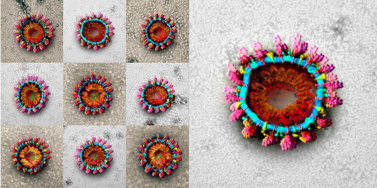
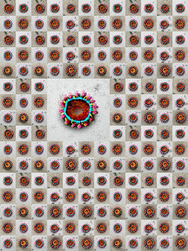
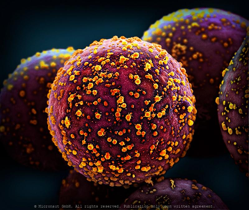
Coronavirus Structures - CoV2
Coronavirus structures. 9 original TEM image scans, hand-colored by Martin Oeggerli.
Coronavirus Structures - CoV2
Coronavirus structures. 9 original TEM image scans, hand-colored by Martin Oeggerli.
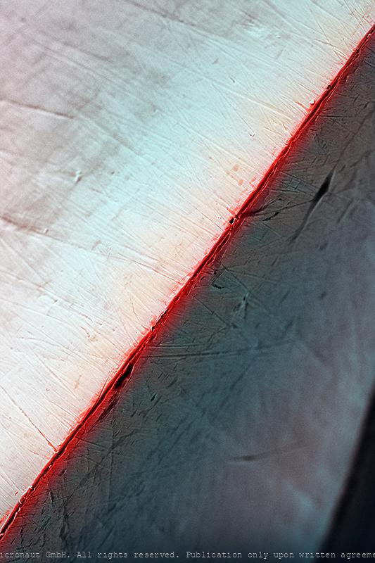
Cutting Edge
Forged Steel of a Guarda Knife (cutting edge) hand colored SEM picture by Martin Oeggerli (coloration) and Theo Bühler (SEM scan).
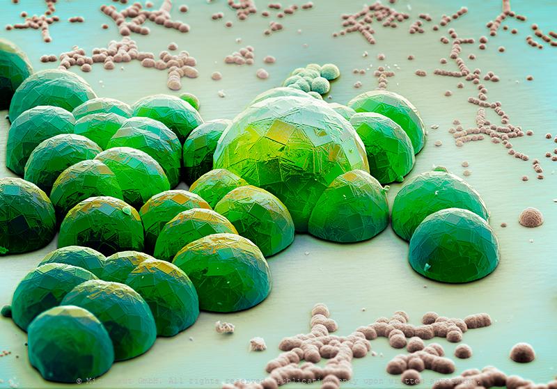
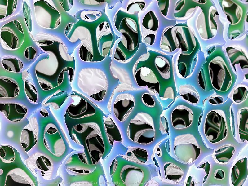
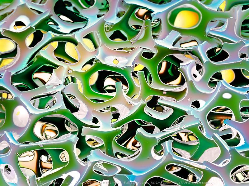
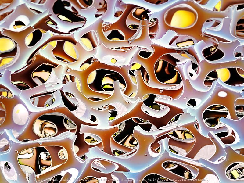
Mammalian Fat Cell
Mammals use specialized connective tissue, called adipose tissue, as cushion, to store energy and to prevent excessive loss of body heat. Technically, adipose tissue is highly organized and consists of 14-sided hexagons. This optimizes the storage capacity of individual fat cells, while reducing material costs and intercellular gaps to the minimum.
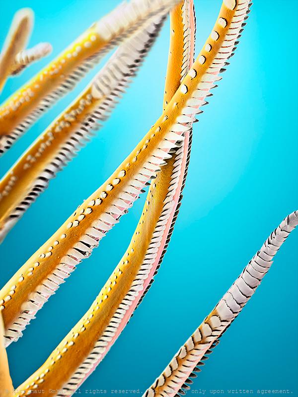
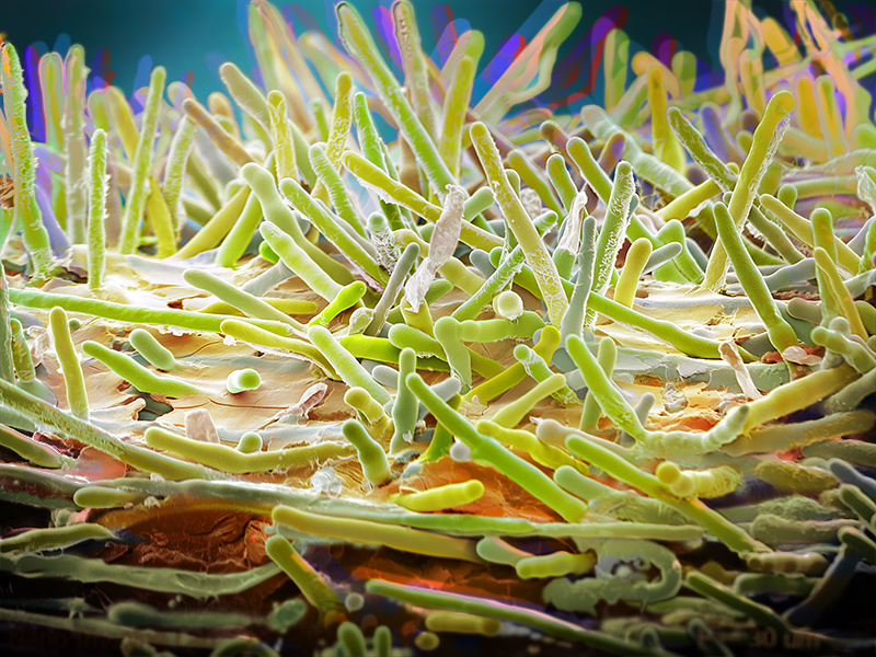
Sensory hairs of a shrimp
Leg hairs (called setae) of a shrimp (crustacea), hand-colored scanning-electron-micrograph by Martin Oeggerli (Micronaut). Most rustaceans (e.g. lobster, crabs, and shrimp) have an extensive array of sensory hairs covering their bodies and they have many different purposes. The setae are the tiny hairs on the legs of the shrimp. Setae can be used to filter food through the water and push it toward the mouth. Some shrimp use the setae to incubate fertilized eggs, and others use setae for grooming. Furthermore, the setae can be used to detect movement in the water, detecting current changes and water flow. It has been reported that some species have also developed highly specialized setae for chemosensory communication. Others use setae to detect water- and substrate-born vibrations and sound as part of their intraspecific communication. For example the sensory hairs (setae) located on the claws of the Australian freshwater crayfish are sensitive to water vibration frequencies between 150-300Hz. This picture shows the tips of setae which are arranged at the very tip of the legs of a freshwater shrimp (unknown species).
E. coli on an LP -(CRISPR-Cas Mediated Genome Editing)
This hand-colored scanning-electron-micrograph (SEM) shows cells of the human gut bacterium Escherichia coli (E. coli) on a phonograph record of Ludwig van Beethoven’s Moonlight Sonata (Sonata No 14 in C). E. coli has been engineered to express the molecular components of another bacterium’s CRISPR-Cas system - an adaptive bacterial immune system that enables it to write a permanent record of the RNAs within the cell into its own genome where it is safely stored. Much like the Beethoven’s composition, this record is preserved over many generations. Scientist can retrieve it at any time to reveal the mysteries of the bacterium’s transcriptional history.

Tip of a hypodermic needle
Tip of a thin surgeon Needle (25g; 1.5; NEO)
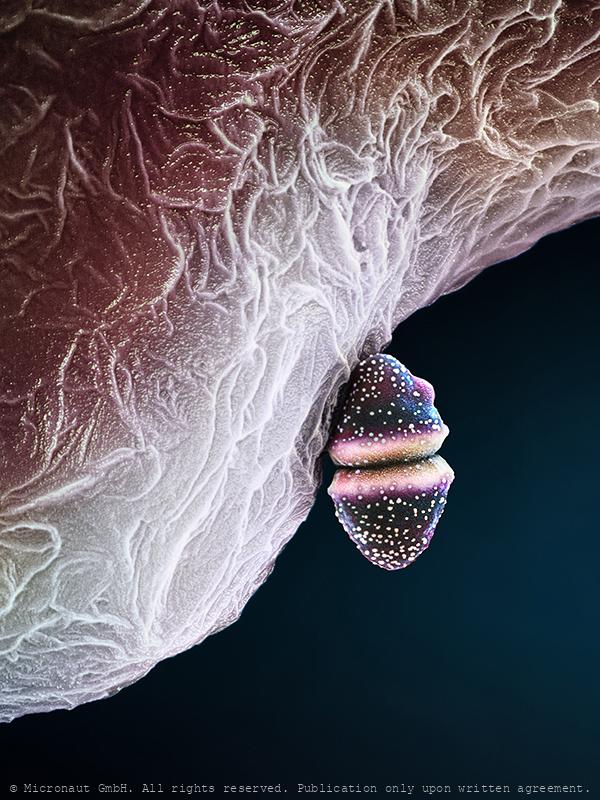
Honey Bee Stinger (Lancet)
This image was produced using scanning electron microscopy in combination with selectively assigning different colors to different structures. By creating a 3D-like photorealistic appearence, the artist aims to expose the delicate fine-structure at the tip of a honey bee (Apis mellifera) stinger. The stinger of a bee is a modified chitinous ovipositor and consists of three parts: a stylus and two barbed slides (or backwards sloping barbed 'lancets'), one on either side of the stylus. In fact, the bee does not have to push the sting in since it is drawn in by the barbed slides which move alternately up and down the stylus so when the barb of one slide has caught and retracts, it pulls the stylus and the other barbed slide into the wound. When the other barb has caught, it also retracts up the stylus pulling the sting further in. This process is repeated until the sting is fully in and even continues after the sting and its mechanism is detached from the bee's abdomen. A sting from a bee (honey bee, bumblebee, sweat bee, etc) can be quite painful. The injected venom or toxin (apitoxin, melittin, histamine, other biogenic amines ) of insects is quite different. Therefore, the body's reaction to a bee sting may differ significantly from one species to another. For about 2 percent of people, a hypersensitivity can develop after being stung, creating a more severe reaction when stung again later. In people with insect sting allergy, a bee sting may trigger a dangerous anaphylactic reaction that is potentially deadly. The sting of a honey bee also releases pheromones that prompt other nearby bees to attack. This alarm process is accelerated if the bee is fatally injured. Although widely believed, a worker honey bee can sting not only once; this partial misconception only happens if the skin of the victim is sufficiently thick and elastic, such as a mammal's. Wasps have stingers with smooth barbs, enabling them to retract the stinger and to sting multiple times. Queen
Weight-in of a cell (H. sapiens)
human Cell on an AFM cantilever. Atomic force microscopy (AFM) is a powerful, multifunctional imaging platform that allows biological samples, from single molecules to living cells, to be visualized and manipulated. The combination of AFM-based imaging and spectroscopy allows 3D manipulation with molecular precision. In this picture, we measure the weight of a cell with with AFM (cantilever). It is anticipated that in the next decade AFM-related techniques will have a profound influence on the way researchers view, characterize and solve fundamental challenges that cannot be addressed with other techniques.
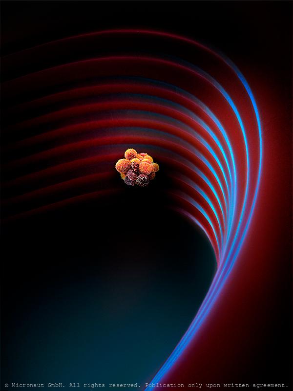
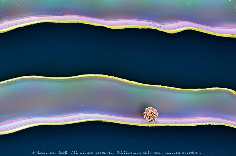
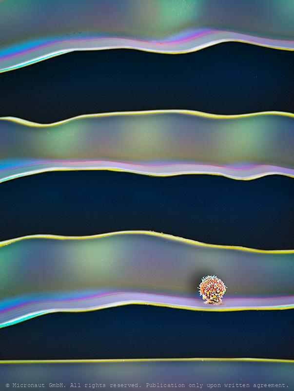
Cancer spreads during sleep (Circulating Tumor Cell Cluster)
Circulating Tumor Cell Cluster (CTC cluster; metastasis) on a microfluidic device
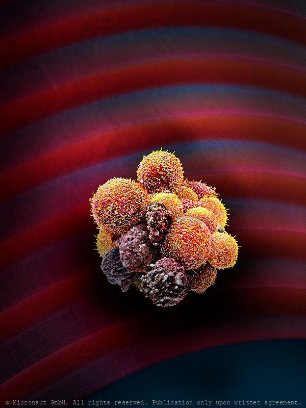
Cancer Spreads During Sleep - Circulating Tumor Cell Cluster on Microfluidic Device
CTC detail at night: The metastatic spread of cancer is achieved by the haematogenous dissemination of circulating tumour cells (CTCs). It is often assumed that CTCs are constantly shed from growing tumours - However, Nicolas Aceto and colleagues have observed a striking and unexpected pattern of CTC generation dynamics in both patients with breast cancer and mouse models, highlighting that most spontaneous CTC intravasation events occur during sleep. By M Oeggerli, I Krol and N Aceto, supported by University Basel, Pathology, University Hosp. Basel, and ETHZ.
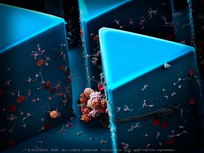
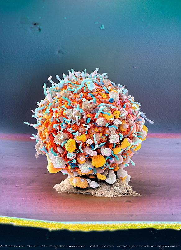
Circulating Tumor Cells, Nr.1
Cancer cells are carried through the bloodstream either as single circulating tumor cells (CTCs) or as multicellular groupings (CTC clusters). In contrast to relatively small single CTCs, the number and dimension of CTC clusters favours the formation of new matastasis sites. This artistically colored scanning-electron-micrograph (SEM) illustrates the new microchip technology which is used to capture and analyse human CTC clusters. By analysing a blood sample of a breast cancer patient with a microfluidic device (blue), blood cells and CTCs can be enrichted and picked and prepared for further examination from the smooth surface of the chips triangle-microstructures. At the center of the image you can see a typical CTC cluster, containing 9-12 tumor cells. Circulating tumor cells are believed to exist in most - if not all - tumor types. Once the process of matastasis has started, the number of circulating tumor cells is continuously increased: i.e. in a 20ml blood sample up to 100 billion blood-cells and 10 CTCs, or CTC clusters can be found. It is important to analyse the morphology and biology of CTC clusters: How do they form? What binds single cells in a CTC cluster? How can CTC clusters speed up metastasis formation? The long-term goal of such research projects is to treat and eliminate CTC clusters in patients, thereby preventing primary tumors to metastasize. -- Klumpen aus zirkulierenden Tumorzellen (CTCs) einer Patientin mit metastasierendem Brustkrebs werden mit Hilfe eines selbst entwickelten Mikrofluidik-Chips (blau) isoliert. Als Vergleichsgrösse dienen rote Blutkörperchen (7.5 µm, rot). © Martin Oeggerli, Micronaut.ch
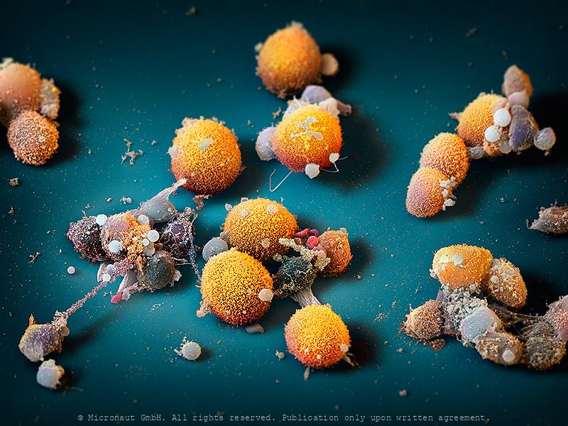
The Plasticity of a Tumor
Artistic coloration of cells of human KRAS-mutated pulmonary adenocarcinoma. The image shows morphologically different cells, as present within the primary lung tumor. Except some granulocytes and blood cells (lymphocytes and erythrocytes; red cells shown in the central part of the image) only tumorigenic cells are shown. The image was produced using scanning electron microscopy in combination with selectively assigning different colors to different structures. By creating a 3D-like photorealistic appearence, the artist aims to expose the complex nature and morphologic plasticity of tumor cells present in a primary tumor.
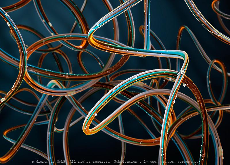
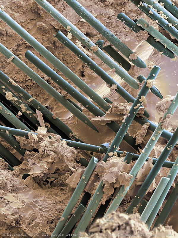
Spider Silk, Nr.1
Spider silk is a protein fibre spun by spiders. Spiders use their silk to make webs or other structures, which function as sticky nets to catch other animals, or as nests or cocoons to protect their offspring, or to wrap up prey. They can also use their silk to suspend themselves, to float through the air, or to glide away from predators. Most spiders vary the thickness and stickiness of their silk for different uses. A single spider can produce up to seven different types of silk for different uses. Consisting of mainly protein, silks are about a sixth of the density of steel (1.3 g/cm3). As a result, a strand long enough to circle the Earth would weigh less than 500 grams (18 oz). Making the silk acidic (pH 4) is a protection from fungi and bacteria that would otherwise digest the protein. The spider silk shown here results from combination of four fibres. This hand-painted picture was produced using a scanning electron microscope, by Micronaut (Martin Oeggerli).
Spider Silk, Nr.2
Spider silk is a protein fibre spun by spiders. Spiders use their silk to make webs or other structures, which function as sticky nets to catch other animals, or as nests or cocoons to protect their offspring, or to wrap up prey. They can also use their silk to suspend themselves, to float through the air, or to glide away from predators. Most spiders vary the thickness and stickiness of their silk for different uses. A single spider can produce up to seven different types of silk for different uses. Consisting of mainly protein, silks are about a sixth of the density of steel (1.3 g/cm3). As a result, a strand long enough to circle the Earth would weigh less than 500 grams (18 oz). Making the silk acidic (pH 4) is a protection from fungi and bacteria that would otherwise digest the protein. The spider silk shown here results from combination of four fibres. This hand-painted picture was produced using a scanning electron microscope, by Micronaut (Martin Oeggerli).
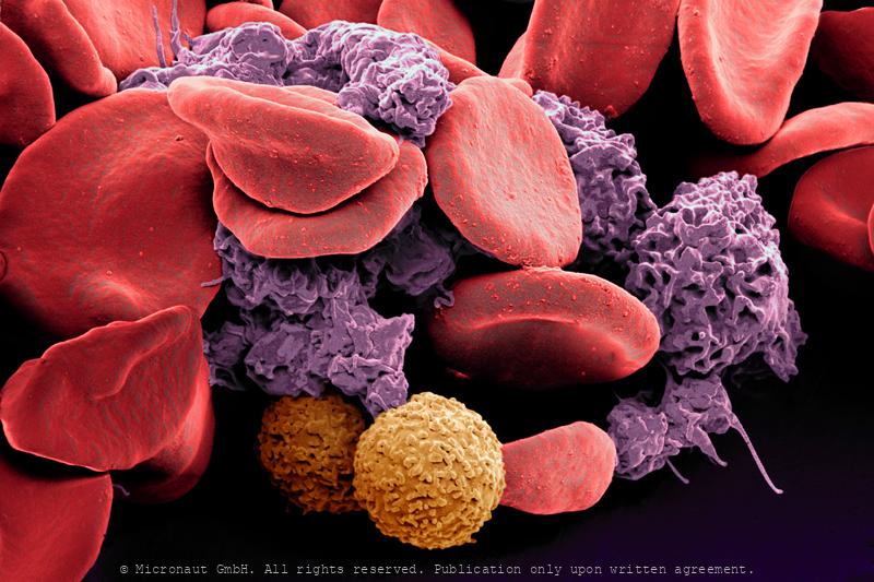
Blood cells
Blood cells
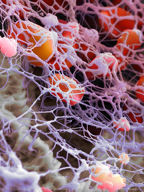
Fibrin network and blood cells (H. sapiens)
Human blood cells 2 minutes after initiation of the blood coagulation process. The fibrin network is already built up. By the way, these are the cells of the author...
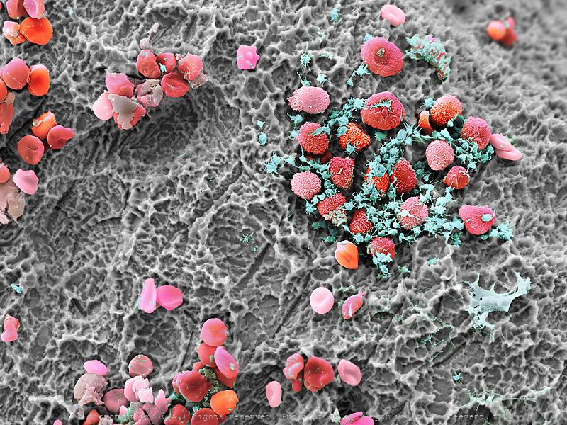
human blood cells during coagulation process
human blood cells during coagulation process
Blood spot (showing various blood cells)
Blood spot (showing various blood cells)
Cellular network
Prostate cancer (in-vitro) cell culture. LNCaP cells are a cell line of human cells commonly used in the field of oncology. LNCaP cells are androgen-sensitive and derive from a human prostate adenocarcinoma (lymph node metastasis) of a 50-year-old caucasian male patient. They are adherent epithelial cells growing in aggregates and as single cells. Prostate cancer is the most frequently diagnosed cancer in men. One major obstacle in prostate cancer research is the lack of cell lines that closely mimic human disease progression. Two hallmarks of metastatic human prostate cancer include the shift of aggressive PCa from androgen-sensitivity to an androgen insensitive state, and the preference of primary prostate cancer cells to metastasize to bone. Although the generation of androgen insensitive cell lines has been demonstrated in “classic” cell lines (DU145 and PC3), the behavior of these cells in bone metastasis does not fully mimic clinical human disease. Therefore, LNCaP sublines have been generated to develop an androgen insensitive prostate cancer cell model that more closely mimics clinical disease.
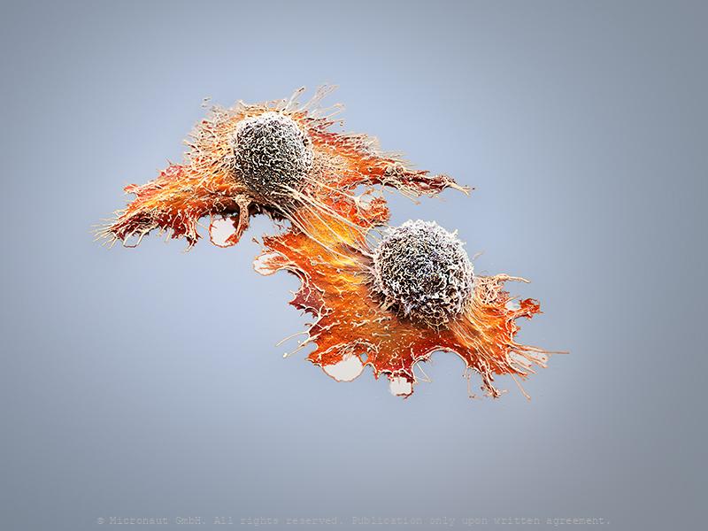
Invasively Growing Cancer Cell
Invasively growing human cancer cell, in vitro. Cancer cells develop the ability to move themselves, sooner or later. The picture shows an invasively growing tumor cell, which is pulling itself around with the help of its appendages. In comparison to a healthy cell its extensions are smaller and because of the quicker assembly and disassembly of the appendixes, it is able to move faster. -- Die Transformation einer gesunden Zelle in eine Tumorzelle ist ein komplexer Vorgang und bedingt eine ganze Reihe verschiedener genetischer Veränderungen. Tumorzellen entwickeln dabei früher oder später die Fähigkeit sich fortzubewegen und Metastasen zu bilden. Das Bild zeigt eine invasive Prostatakarzinomzelle, welche häufig Metastasen im Knochenmark bilden. Mit Hilfe der Fortsätze zieht sich die Zelle über die Unterlage. Im Vergleich zu einer gesunden Zelle bildet diese invasive Tumorzelle deutlich kleinere Fortsätze aus und ist dank schnellerem Auf- und Abbau (der Fortsätze) in der Lage, sich wesentlich schneller fortzubewegen.
Clonal evolution versus cancer stem cell theory
In cancer research two fundamentally different mechanisms explaining tumor progression currently split the positions: Clonal evolution versus cancer stem cells. To develop more effective cancer therapies it will be critical to determine which theory holds true. If most cells can proliferatie and metastasize, then virtually all cells must be eradicated to cure the disease, whereas the specific elimination of cancer stem cells would be sufficient, if the theory of clonal evolution is a myth...
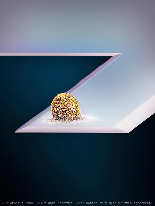
Cancer Stem Cell (DLD1)
Colored Scanning Electron Micrograph of a Colon Cancer Stem Cell (DLD1): cell culture of a non-adherent cancer stem cell line derived from a primary colon tumor.
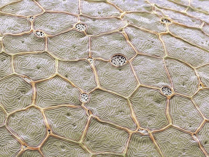
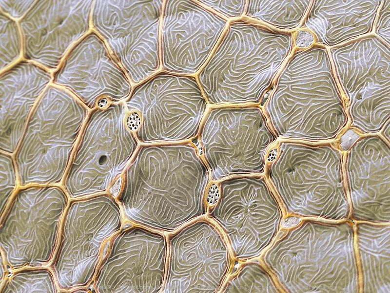
Comparison of two flower morphologies present in Asteraceae
Comparison of two florets from a compound flower (The Common Daisy; Bellis perennis). Florets from the border of the influorescence (left) have developped a special morphology with five long clean and seamlessly adnate white sepals, in order to attract pollinators, whereas florets from the center have totally reduced sepals. Central florets are densely packed and mainly required for increased pollen- and seed production.
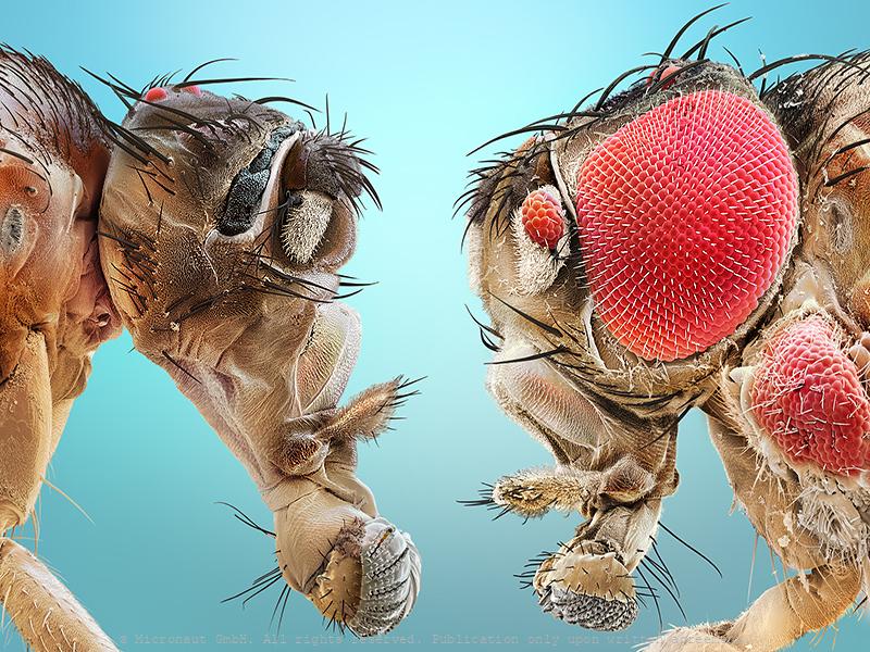
Uneven brothers (D. melanogaster), Nr.3
Coloured scanning electron micrograph (SEM) of two mutant Fruitfly (Drosophila melanogaster) heads. Wildtype (i.e. normal) Fruitflies have two compound eyes (red) - one on either side of the head. Small bristles between the single lenses of the eye make sure it cannot be covered with dust or dirt particles. Genetically manipulated Flies can either lack compound eyes completely (left) or have additional eyes on the antennae, legs and other body areas (right). Fruit flies are widely used in genetic experiments, particularly in mutation experiments, because they reproduce rapidly and their genetic systems are well understood. This image visualizes how easily the results of the genetic modification can be observed in the Fruitfly which is one of the main reasons why it is still the most frequently used model organism in genetics despite more than 100 years of experimental research. left: Sine oculis-1 (so1) variant which lacks the compound eyes completely. right: Eyeless variant with ectopically expressed compound eyes under dpp-promotor in all imaginal discs. Handkoloriertes Raster-Elektronen-Mikroskopiebild zweier Fruchtfliegen-Mutanten. Die Taufliege oder Fruchtfliege (Drosophila melanogaster) ist das klassische Untersuchungsobjekt in der Genetik und Entwicklungsbiologie. Wildtypen, d.h. genetisch unveränderte Fruchtfliegen, besitzen zwei grosse rote Komplexaugen - je eines auf jeder Seite des Kopfes. Die Komplexaugen werden durch Borsten zwischen den winzigen Einzelaugen vor Verschmutzung und Staub geschützt. Die Entwicklung des Auges wird während der Embryonalentwicklung Larve durch bestimmte Gene gesteuert. Durch Veränderungen (Mutation) von Genen lassen sich z.B. Position oder Morphologie der Komplexaugen gezielte beeinflussen. Im Vergleich zu einem Säugetier ist die Fruchtfliege genetisch viel einfacher aufgebaut und weist lediglich vier Paar Chromosomen auf. Diese Tatsache hat, neben der raschen Generationsfolge und einfachen Zucht, wesentlich dazu
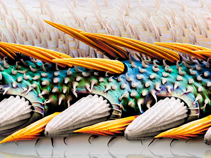
The Leading Edge - Aerodynamic key parameter of the fly wing
The flight of insects has fascinated physicists and biologists for more than a century. Yet, until recently, researchers were unable to rigorously quantify the complex wing motions of flapping insects or measure the forces and flows around their wings. From an aerodynamics point of view, the wing leading edge (which is shown in the picture) fundamentally affects the way we understand how insect-like wings at low Reynolds number work.
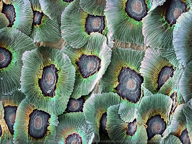
The scales on butterfly wings derive from highly complicated flat hairs. In certain species, they lead to iridiscent color by breaking the sunlight into different wavelengths.
The scales on butterfly wings derive from highly complicated flat hairs. In certain species, they lead to iridiscent color by breaking the sunlight into different wavelengths.
The scales on butterfly wings derive from highly complicated flat hairs. In certain species, they lead to iridiscent color by breaking the sunlight into different wavelengths.
The scales on butterfly wings derive from highly complicated flat hairs. In certain species, they lead to iridiscent color by breaking the sunlight into different wavelengths.
Gyroid structures (Parides sesostris)
The majority of butterfly colors are pigmentary. However, scientists have now found out that vibrant green wing colors can also be induced by complex three dimensional crystals - called gyroids (see porous red patches underneath corrucated green scale surface). The gyroid network forms during the butterflies' cocoon phase and refracts light in the adult insect's wing. Since the structure measures only a few hundred nanometers it remains difficult to document. For National Geographic, Micronaut has teamed up with Marco Cantoni from the 'Ecole Polytechnique Federale De Lausanne' to visually explore and document the gyroid arrangement in the wing of Parides sesostris, the Emerald-patched Cattleheart Butterfly. The resultant image shows a cross-section through a single wing scale and has been produced with a 'Focused-ion beam' (FIB) electron microscope. The technology uses a beam of focused ions to manipulate the microscopically small sample and the instrument is usually applied in material investigations. Similar to the 'Scanning-Electron-Microscopes' (SEM), the FIB also allows investigations with extremely powerful magnification.
Distinctive Masterpiece
Bild im Querschnitt; Painting disection
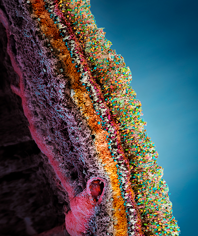
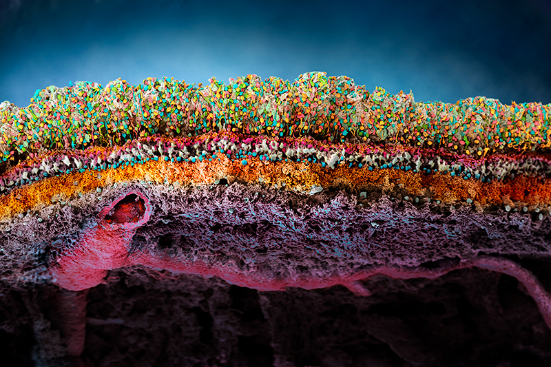
The human Retina - Nr. 2 (2019), horizontal
With far more than 100 million nerve cells, the retina is the first stage of our visual system and our window to the outside world. The process includes detection of light impulses (photons) by different light receptors, in general called rods (120 million cells) and cones (6 million cells), and the fast and continuous translation, filtration and post-procession into electrical signals (or nerve impulses). These signals are then passed through the optical nerve’s 1.5 million fibres to the visual centre of the brain and reinterpreted into a cohesive image.The flexibility and economy with which retinal cells work together remains beyond our powers of imagination: the eye reports numerous signals to the brain at once, including separate detection of light (light on) and dark (light off), general patterns and finest details, movements and different hues (including: red, green and blue). This literally makes the eye a camera with 10 to 15 different films and despite it appears so naturally to us, the perception of an image is the result of a most complex interaction process involving millions of cells and the electrical signals they produce and pass on to the brain. What you can see on this image is the (1) rods and cones layer. It is located below the pigment epithelia which has been cut away during the preparation and is not visible on this image (black space on top). Below the rods and cones layer you can find the (2) outer nuclear layer (ONL) which contains the nuclei of the rods and cones. The adjacent (3) outer plexiform layer (OPL) is followed by the (4) inner plexiform layer (IPL), which contains numerous cell types, including horizontal cells, bipolar cells and amacrine cells. At the very bottom you can see the (5) inner nuclear layer (INL), followed by the (6) ganglion cell layer with two (7) blood vessels shown at left and (8) nerve fibers (which form the optical nerve).
Skin & Bacteria
Human skin bacteria play an important role in the production of human odours. There is a strong correlation between human body odour and the species composition of skin bacteria. Freshly secreted human sweat is odourless. Staphylococcus epidermidis, Pseudomonas aeruginosa, Corynebacterium minutissimum, Brevibacterium epidermidis, Bacillus subtilis are commonly found on the human skin and are associated with differences in body odour production. Interestingly, scientific studies show that the composition of human skin microbiota leads to different odours and thereby determines the attractiveness of humans to (malaria) mosquitoes (Verhulst et al, PlosONE, 2010).
Hirudino medicinalis surface
Hirudino medicinalis surface
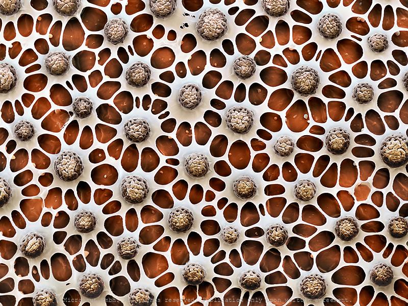
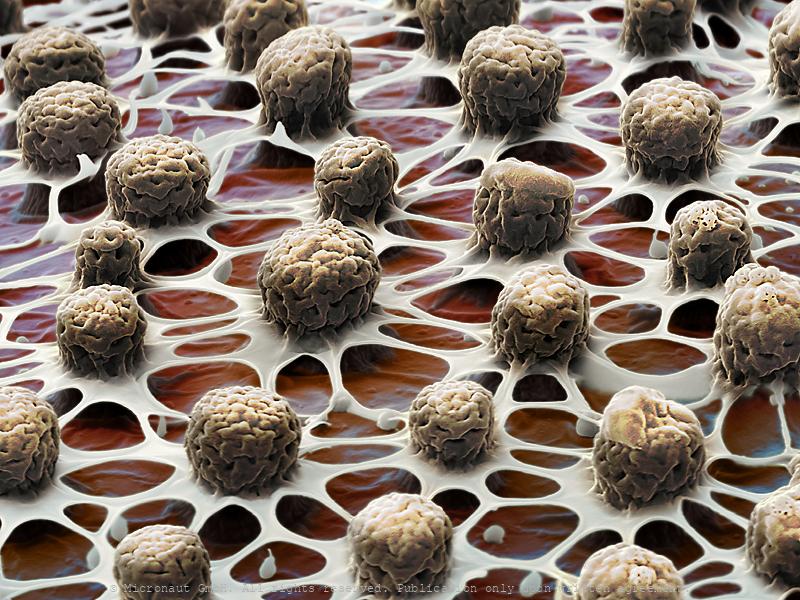
The Truffle (Tuber spp) microbiome
Truffles (i.e. the Périgord truffle Tuber melanosporum or the white truffle Tuber magnatum), Owing to their unique aromas, truffle fungi (Tuber spp.) are regarded as most precious food delicacies. Since many years, scientists try to decrypt aroma production in truffles. Over the last decade, Prof. Richard Splivallo has explored along with international colleagues the genomes of various truffle species as well as the specific contribution of bacteria in truffle aroma formation. These scientists could show that, based on the genomes, truffles seemed to possess most of the molecular machinery needed to produce many of their flavor compounds. Yet, Prof. Splivallo and colleagues could also show that bacteria colonizing truffle fruiting bodies were responsible for the production of key truffle volatiles. This illustrates that overall truffle aroma results from the intimate interaction of truffles with bacteria that are trapped inside of truffles. Manually colored scanning-electron-micrograph (SEM) by Martin Oeggerli, kindly supported by Richard Splivallo. The picture shows isolated bacteria from a truffle (Tuber melanosporum).
Intestinal Bacteria
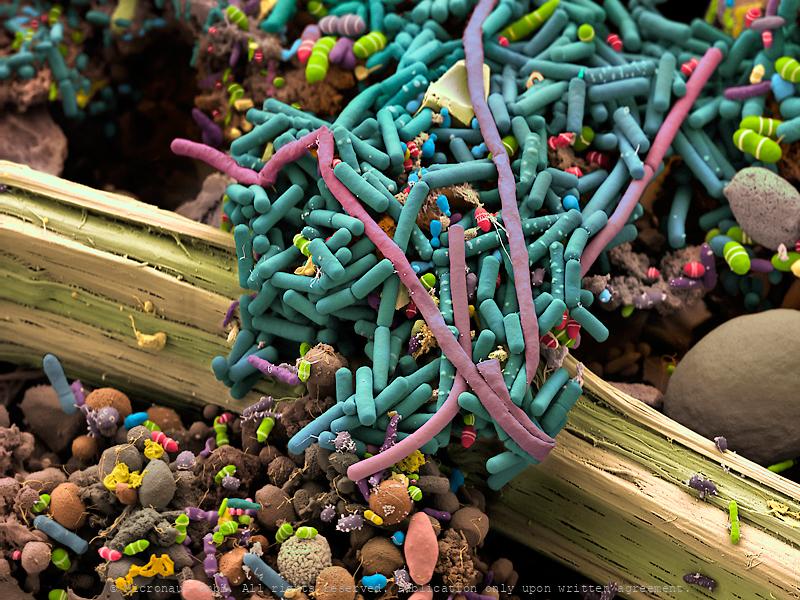
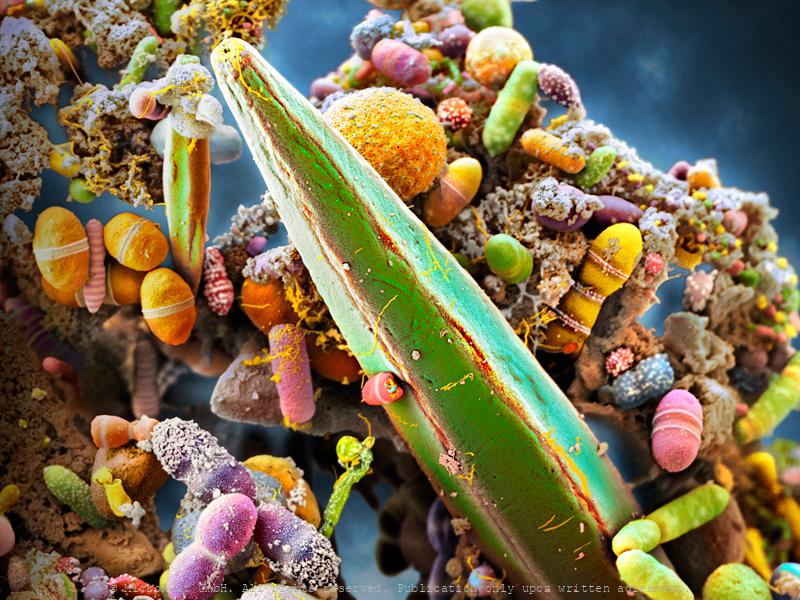
Human excrements with intestinal bacteria. The human gut teems with bacteria (streptococci, staphylococci, enterococci, enterobacteria, mycobacteria, spirochetes, mycoplasma, corynebacteria clostridia, or lactobacilli). Many of their species are still unknown. They help us digest food and absorb nutrients, and they play a part in protecting our intestinal walls. Gut bacteria may also help regulate weight and ward off autoimmune diseases. In adult humans, the intestine bacterial flore makes up about five percent of the body weight, forming a fragile ecosystem sensitive to stress, unhealthy food, alcohol or illness. Under unfavorable circumstances our health is directly affected and as a result we suffer from e.g. overweight, mood changes or prone to various diseases. In this sample, numerous intestinal bacteria can be detected. Additionally, a plant fiber has passed the intestinal tract unharmed and spans across the frame (center). On the right hand side (half-cut) the relatively large and spherical shaped cyste of a Giardia parasite (brown) is visible. Apart from this unusual parasitic supplement, the documented bacterial fauna looks fairly diverse and well-sorted…
The Human Microbiome, Nr.1
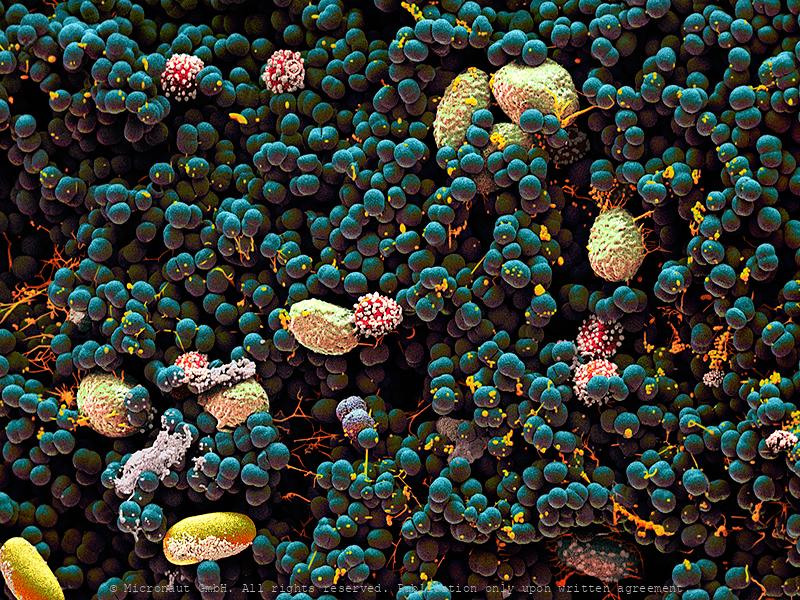
Artistic coloration of human excrements, that contain billions of intestinal bacteria, including Epulopiscium sp. - the world’s largest bacteria species! Artistic coloration of the microbiome present in a healthy human gut, containing billions of intestinal bacteria, including Epulopiscium sp. - the world’s largest bacteria species! The human gut teems with bacteria: streptococci, staphylococci, enterococci, enterobacteria, mycobacteria, spirochetes, mycoplasma, corynebacteria, clostridia, and lactobacilli. Many of their species are still unknown. They help us digest food and absorb nutrients, or play a role in protection of intestinal walls. Many gut bacteria further regulate weight and ward off autoimmune diseases. In adult humans, the intestinal flora is responsible for up to five percent of the body weight, forming a fragile ecosystem (the human microbiome) which is sensitive to: stress, unhealthy food, alcohol or illness. Under unfavorable circumstances our health is directly affected by bacteria that live inside of us, and as a result we may suffer from: overweight, mood changes or become prone to various diseases. In the central part of the image you can see the world’s largest bacteria - Epulopiscium sp. which spans between 200 – 700 µm, i.e. 350-times bigger than E. coli, or B. subtilis. Epulopiscium was first discovered in 1985 in the intestines of a surgeonfish, where it is only active during the day and believed to control the pH within the gut of its hosts. Because of its huge size, the giant exhibits many unusual characteristics, including a very special ‘rhomboid’ morphology. This image has been produced using scanning electron microscopy (SEM) in combination with selectively assigning different colors to different bacteria species and structures. By creating a 3D-like photorealistic appearence, the artist aims to expose the diversity of bacteria that can be found within human feces.
Belly Button Bacteria
Belly Button Bacteria and Fungi Spores under the scanning electron microscope. Our body is home for lots of microbial species and interestingly, they have not been investigated until very recently. We imagine microbes as something bad, but that's not the case. In fact, most microbes on our body are either good for us or simply present, whether on your toe or in your nose. Right now on our bodies, bacteria are fighting other bacteria, fungi and even viruses. Among your skin’s true warrior clans are species of the genus Bacilllus such as Bacillus subtilis. Bacillus subtilis produces antibiotic compounds that can kill other bacteria and even foot fungi. Right now it may be doing this on your skin. We tend to think of the life on our skin as somehow stable and yet like any wild kingdom the individuals alive at any moment are the result of millions of independent wins and losses. Scientists have recently identified the Staphylococci, Corynebacteria, Actinobacteria, Clostridiales, and Bacilli as the most common groups of bacteria on our bodies. Some can be beautiful: - Species of Micrococcus (M. luteus) produce yellow colonies, others (M. roseus) red ones, at least when grown in agar. Micrococcus species are aerobes and can deal with extreme drought and long periods of starvation. This toughness predisposes them to a life spent on our desert-dry flesh. - The Clostridiales include bacteria like Anaerococcus and Clostridia. Despite some of them can be bad news, most are harmless or even beneficial. The Clostridiales are spindle-shaped and motile anaerobes. They may be growing in your belly button, or maybe inside your ear. - Like almost any bacteria in the wrong place, Staphylococcus species such as Staphylococcus epidermidis can become pathogens but when living on your skin, Staphylococcus are far more likely to be beneficial, working as our first line of defense against pathogen invasion.
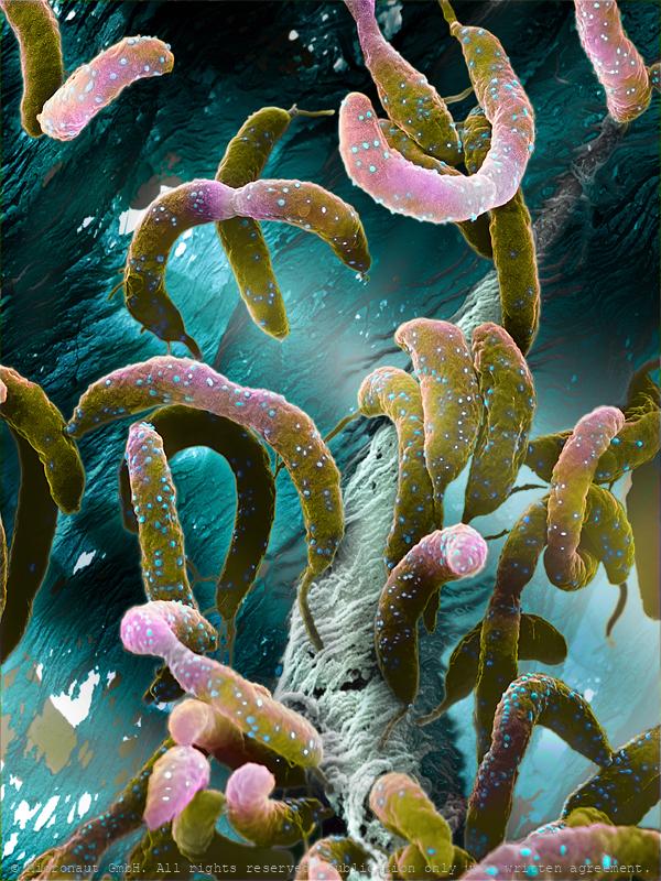
Waterbacteria (Caulobacter crescentus)
Water bacteria exist in two different forms: a mobile cell (called a “swarmer cell”) which is equipped with a hair-like flagellum to swim freely in the water and an immobile cell (called a “stalked cell”) which is equipped with an adhesive to attach the cell to the surface. Interestingly, both cells arise from asymmetrical cell division which is a complex and exceedingly rare process in nature and can e.g. lead to cancer. Waterbacteria allow scientists to study asymmetrical cell division in depth and to learn more about the process of cell division in general. Additionally, the stalked cell produces THE strongest glue in nature, for a safe anchorage to any surface and induces the formation of biofilms. It’s a nasty problem for the maritime industry, since biofilms increase drag and annually lead to additional costs and waste of fuel. Best scientific image 2008. -- Caulobacter teilt sich asymmetrisch in zwei Tochterzellen mit unterschiedlichem Entwicklungsprogramm. Die Bakterienart kann dadurch besser auf sich ändernde Umweltbedingungen reagieren. Forschungsversuche an Wasserbakterien dienen längerfristig dazu zelluläres Verhalten zu berechnen und einfache lebende Systeme zu konstruieren. Ausgezeichnet als Bestes Bild der Forschung 2008 (1. Rang Kategorie Faszination Forschung).
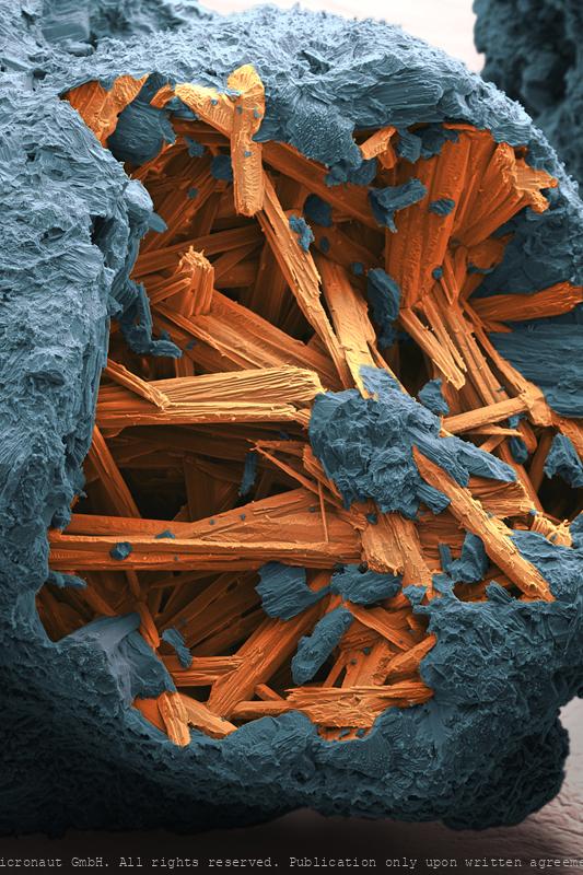
Doxycycline - Antibiotics
Antibiotics (Doxycycline)
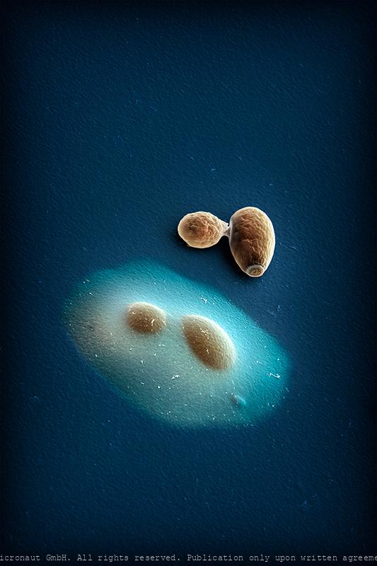
Encapsulated vs. normal yeast cells
Budding yeast (Saccharomyces cerevisiae); hand-colored scanning-electron-micrograph by Micronaut (Martin Oeggerli). Modification of cell surfaces with synthetic hydrogels is a promising approach for controlling cell behaviour. Here, Vanella and colleagues developed a genetically controlled system that enables cells to auto-encapsulate inside protective hydrogel capsules. The image shows an encapsulated budding yeast cell directly adjacent to a non-encapsulated one. This system links inducible gene expression to a synthetic capsule that protects target cells against lysis and mediates communication with the extracellular space through a permeable hydrogel.
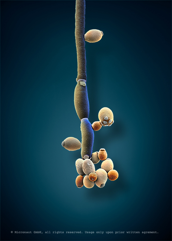
Face/Off - The interchangeable fungi (Candida albicans)
Candida albicans (laboratory culture), hand-colored SEM scan by Martin Oeggerli (Micronaut). Candida albicans is the most common member of the human gut flora and it can also survive outside the human body. In healthy human adults, it is a commensal microbe of the gastrointestinal tract and mouth in 40–60%. However, C. albicans can also become pathogenic in immunocompromised individuals, causing the human infection candidiasis, which results from an overgrowth of the fungus and can often be observed in e.g. in HIV-infected patients. Candidiasis is causing 2800-11200 deaths in the US yearly. Furthermore, C. albicans is the most common fungal species isolated from biofilms. It is also used as a model organism to study fungal pathogens. According to new studies, C. albicans can cross the blood–brain barrier in mice. Interestingly, C. albicans can grow as both, yeast (opaque and GUT form) and filamentous cells (pseudohyphal form): due to limited mobility, fungi have evolved mechanisms to adapt to sudden chemical and/or physical variation in their environment and scientists are using C. albicans to study eukaryotic responses to environmental changes by switching morphologies from yeast to hyphae growth.
Scientists & Diamonds (Nr. 2)
Everybody knows that diamonds are beautiful and ever-lasting jewels symbolising purity and commitment. What might surprise you is that the heavenly purity of diamonds can be used in quantum technology to reach unprecedentedly high performances. Ultrapure diamonds play an important role for the development of hi-tech measuring devices in medicine and computer science in the future. Quantum technology builds on quantum theory, which describes the behaviour of microscopic objects such as atoms and photons, which is different from that of objects that we observe in our daily life. Over the years, quantum technology has led to amazing innovations (computers, the internet and much more) that to a large extent shape today’s society. The next step will be to develop methods for measuring and controlling individual atoms and photons: no wonder that work in this area won the 2012 Nobel Prize in Physics! The picture shows a hand-colored SEM picture of a diamond nano cantilever attached to a device to handle and transport the tip. Oeggerli slowly breathes life into his works by painstakingly selecting and masking different structures with color, layer upon layer utilizing his laptop touchpad. The process allows him to set focus on the mysterious shape of a nano-diamond AFM tip.
Scientists & Diamonds (Nr. 1)
Everybody knows that diamonds are beautiful and ever-lasting jewels symbolising purity and commitment. What might surprise you is that the heavenly purity of diamonds can be used in quantum technology to reach unprecedentedly high performances. Ultrapure diamonds play an important role for the development of hi-tech measuring devices in medicine and computer science in the future. Quantum technology builds on quantum theory, which describes the behaviour of microscopic objects such as atoms and photons, which is different from that of objects that we observe in our daily life. Over the years, quantum technology has led to amazing innovations (computers, the internet and much more) that to a large extent shape today’s society. The next step will be to develop methods for measuring and controlling individual atoms and photons: no wonder that work in this area won the 2012 Nobel Prize in Physics! The picture shows a hand-colored SEM picture of a diamond nano cantilever attached to a needle to put the chip in place. Oeggerli slowly breathes life into his works by painstakingly selecting and masking different structures with color, layer upon layer utilizing his laptop touchpad. The process allows him to set focus on the mysterious shape of a nano-diamond AFM tip.
Crystalline Diamond Pyramids
Single crystalline diamond pyramids for applications in nanoscale magnetic field sensing Color centers in diamond are optically active point defects in the crystal lattice. In the Quantum Sensing Group of Patrick Maletinsky at the University of Basel, such color centers are used as sensors to perform magnetometry with single-spin sensitivity and nanoscale spatial resolution. To harness the full potential of such sensors a high degree of control over the positioning of the point defect with respect to the sample surface is essential. In a novel approach to fabricate the next generation of color center-based sensors, top-down fabrication is combined with bottom-up diamond overgrowth. The depicted diamond is a result of such a test series where cylindrical diamond pillars were overgrown to form pyramids. Oeggerli slowly breathes life into his works by painstakingly selecting different structures with different colors, layer upon layer on his laptop. The process allows him to set focus on the mysterious structures of the invisibly small scientific experiments and structures.
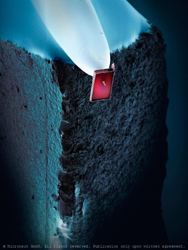
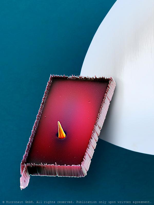
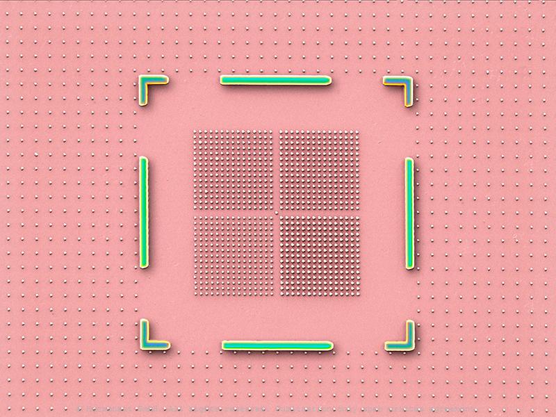
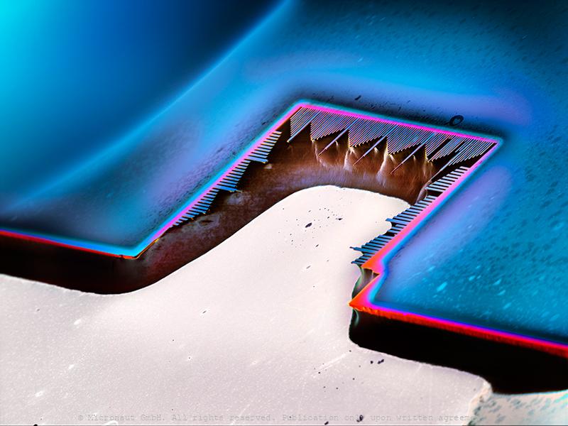
The Diamond Chip
Structured single crystalline diamond chip during the fabrication process of magnetic field sensors Magnetic field sensors based on color centers in diamond are at the forefront of emerging sensing technologies for high sensitivity magnetometry at the nanoscale. These sensors are investigated in the Quantum Sensing Group of Patrick Maletinsky at the University of Basel and fabricated by using a top-down approach. Starting with a single crystal diamond chip they are formed by using several subsequent lithography and etching steps. The image shows an intermediate step during the fabrication process, where platelets are etched into the diamond. These are connected to a frame by thin bridges and can be identified by the coordinate system. In total an area of 900 µm x 900 µm is structured like this within one fabrication process. To finalize the sensor elements photonic structures are etched on the platelets before they are finally released from the diamond. Oeggerli slowly breathes life into his works by painstakingly selecting different structures with different colors, layer upon layer on his laptop. The process allows him to set focus on the mysterious structures of the invisibly small scientific experiments and structures.
The Resonator
Hybrid spin-mechanical systems, formed by single spins coupled to mechanical resonators, have attracted ever-increasing attention over the past few years, triggered largely by the prospect of employing such devices as high-performance nanoscale sensors or transducers in quantum computing networks. In the Quantum Sensing Group of Patrick Maletinsky at the University of Basel, such hybrid systems comprising diamond mechanical cantilevers with embedded defect center spins are investigated as depicted in the image. Thereby crystal strain occurring upon cantilever displacement affords a natural and intrinsic mechanism to couple both systems. The research ultimately aims at investigating the potential of these systems for future fundamental physics studies and applications in sensing or information processing.
Synthetic Diamonds
Synthetic Diamonds A natural diamond is made from carbon and is the hardest natural known substance on earth. Natural diamonds are created over a period of one to three billion years, at least 85 miles below the earth’s mantle under natural conditions of very high pressure and high temperature. Once a diamond has been created in these underground conditions, it travels via molten rock to the earth’s surface, where it is mined, refined, and turned into beautiful jewelry or used for industrial purposes. Natural diamonds are slightly more durable, ranking 10 out of 10 on the Mohs Hardness scale, whereas synthetic (lab-) diamonds rank 9.25.
Nano Diamonds
Waterproof (fabric)
Waterproof yet breathable fabric. Material is based on thermo-mechanically expanded polytetrafluoroethylene (PTFE) and can be used in a wide variety of applications such as high performance fabrics, medical implants, filter media, insulation for wires and cables, gaskets, and sealants. The fabric is best known for its use in protective, yet breathable, rainwear.
Niacinamide Nr.2
Niacinamide
Niacinamide, highly enlarged. This B3 vitamin is applied in pharmaceuticals as well as in food and feedstuffs
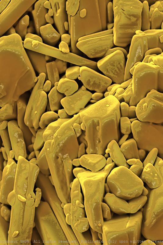
Natriumdicyanamide
Natriumdicyanamide
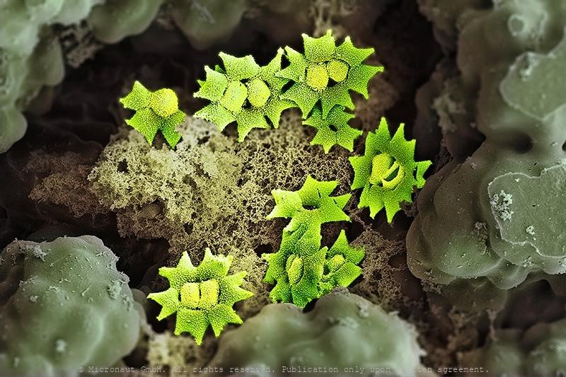
Mosquito egg surface
Adult females lay their eggs in standing water, which can be a salt-marsh, a lake, a puddle, a natural reservoir on a plant, or an artificial water container such as a plastic bucket. Usually many eggs are clumped together (i.e. 'egg rafts'). Each egg has a hydrophobic surface: a thin layer of air is trapped by 'tent-like' micro-structures, to avoid sinking of the egg/egg-raft.
Bone Structure (Mus musculus)
This portion of the Petrous bone (Mus musculus) is protecting the inner ear structure. Though it is one of the hardest, densest bones in the body. Petrous comes from the Latin word petrosus, meaning “stone-like, hard”.
Polyurethan PUR
Polyurethan; hand colored SEM image
Polyurethan PUR, Nr.2
Polyurethane (PUR) is any polymer composed of a chain of organic units joined by carbamate links. -- High-Tech Schaum (Polyurethan; PUR)Viskoelastischer PUR-Schaum, hergestellt nach Bayer MaterialScience Technologie, ist ein offenzelliger, flexiblerPolyurethanschaum mit einzigartigen physikalischen Eigenschaften, die es ihm erlauben, formgetreue Konturen auszubildenund Drücke zu verteilen.Vergrößerung: 125:1 bei Druck auf A6.

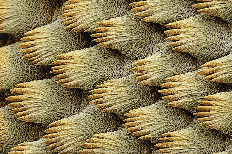
High-Speed Swimsuit - Skin of a Mako Shark
Highly magnified hand-colored scanning-electron-micrograph of Mako (Isurus oxyrinchus) shark skin. Sharks have a covering of dermal denticles that protects their skin from damage and parasites, in addition to improving their fluid dynamics. Single striatet scales (i.e. 'riblets') are overlapping like roof tiles. Each riblet produces microscopically small turbulences, creating a very thin film of water turbulences around the sharks body to reduce lateral drag. This enables Mako sharks to swim as fast as 70 kilometres/h. This effect is only efficient for fast swimming and similar structures are used for submarines, boats and airplanes.

Pitcher plant (Nepenthes hemsleyana), Nr.1
Hand colored scanning-electron micrograph by Martin Oeggerli (Micronaut), showing the slippery surface inside a pitcher plant. Nepenthes hemsleyana is a tropical pitcher plant endemic to Borneo, where it grows in peat swamp forest and heath forest. It appears to rely on a prey trapping strategy unlike other pitcher plants: Interestingly, N. hemsleyana seems to have co-evolved with Hardwicke's woolly bats (Kerivoula hardwickii) which commonly roost in the upper pitchers. Studies shows that the carnivorous plant attracts bats by possessing modified pitfall taps that increase the reflectivity of echolocation calls - bats benefit by finding roosting sites, and the plants gain by receiving nitrogen from bat guano. Unlike in closely related pitcher plants, the upper pitchers of N. hemsleyana feature an expanded waxy zone (this structure is shown in the image) and a watery, less viscoelastic pitcher fluid. The pitchers also appear to lack UV patterns and produce less nectar and odour attractants, which is not required to host the bats.
Zebrafish scales (Danio rerio), Nr.1
Zebrafish scales. Coloured scanning electron micrograph (SEM) of zebrafish scales (Danio rerio) scales. Fish scales are are classified by shape. As the fish grows, the scale increase in size and growth rings called circuli become visible. During cooler months of the year the scale grows more slowly and the circuli are closer together leaving a band called an annulus. By counting the annuli it is possible to estimate the age of the fish.
Zebrafish scales (Danio rerio), Nr.2
Zebrafish scales. Coloured scanning electron micrograph (SEM) of zebrafish scales (Danio rerio) scales. Fish scales are are classified by shape. As the fish grows, the scale increase in size and growth rings called circuli become visible. During cooler months of the year the scale grows more slowly and the circuli are closer together leaving a band called an annulus. By counting the annuli it is possible to estimate the age of the fish.
The epidermis of a tillandsia is covered with sucking hairs. The plant has reduced roots and is specialised on using fog as the primary source of water.
As water becomes a more and more scarce, it's interesting to look around and learn how nature is dealing with the exact same problem, and it reveals interesting techniques and structures. While some plant species such as e.g. Eucalyptus are covering the entire leaf area with a thick layer of wax, the Olive tree has developed flattened hairs on the leaf surface which resemble umbrellas to provide some extra shade for the leaf surface below. Yet the Tillandsia went one step further: the small epiphytic plant which has no roots and virtually no access to groundwater resources, uses special trichomes (which are called sucking hairs) to absorb water from moist air. The picture shows how trichomes cover the entire leaf area. It needs a unique solution to survive in an acrobatic environment.
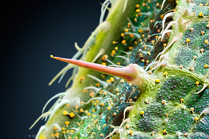
Stinging Nettle hair (Urtica dioica)
The Stinging Nettle (Urtica dioica) is a plant with soft green leafs that are equipped with glands as well as short non-stinging and special elongated stinging hairs (center), from which the species derives its common names: stinging nettle, burn nettle, burn weed, or burn hazel. Stinging hairs (trichomes) produce a painful stinging sensation by injecting a chemical mixture when touched by humans or other animals. They act like hypodermic needles: after the tip breaks off, a chemical mixture composed of histamine, acetylcholine, 5-HT (serotonin), moroidin, leukotrienes and formic acid is injected and causes pain or paresthesia. Furthermore, the plant has a long history of use in medicine, as food source (tea) and as source of fibre.
Material
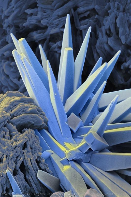
Calcium fluoride
Calcium fluoride is an insoluable ionic compound of calcium and fluorine. It is commonly used as a window material for both infrared and ultraviolet wavelengths, since it is transparent in these regions (0.15-9 micrometers).
Polymeric Filtration
Haute couture supramolecular modification of polymeric filtration membranes allows the stable yet reversible immobilization of tailor-made biocatalysts on the membrane surface. The chemical strategy described in their Communication (DOI: 10.1002/anie.201507020) by P. Shahgaldian et al. is based on multiple-point interactions between the membrane surface on which cyclodextrins have been immobilized and a soluble enzyme–polymer conjugate containing adamantane units.
Catalyst I
Catalyst I
Catalyst
Catalyst
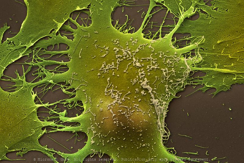
Mammalian cell
Rat cell
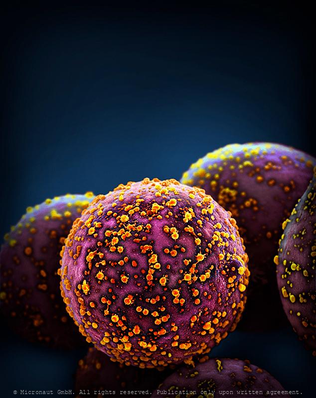
Beads with Viruses
Beads with viruses
Beads with Viruses
Beads with viruses
Superhydrophobic surface (Salvinia molesta)
Aquatic organisms possess structured surfaces which enable them to effectively retain air films while submerged in water. The transfer of these optimised hydrophobic structures on technological surfaces would open up a multitude of applications.
Porcine retina (Sus domesticus)
Porcine Retina (Sus domesticus) With far more than 100 million nerve cells, the retina is our first stage of the visual system and window to the outside world. The picture highlights the neuronal layers of a porcine retina. Seungkuk Ahn and his colleagues have electromagnetically guided viruses to specifically transduce each of the retina layers. The process inside the retina includes detection of light impulses (photons) by different light receptors, in general called rods (120 million cells) and cones (6 million cells), and the fast and continuous translation, filtration and post-procession into electrical signals (or nerve impulses). These signals are then passed through the optical nerve’s 1.5 million fibres to the visual centre of the brain and reinterpreted into a cohesive image.The flexibility and economy with which retinal cells work together remains beyond our powers of imagination: the eye reports numerous signals to the brain at once, including separate detection of light (light on) and dark (light off), general patterns and finest details, movements and different hues (including: red, green and blue). This literally makes the eye a camera with 10 to 15 different films and despite it appears so naturally to us, the perception of an image is the result of a most complex interaction process involving millions of cells and the electrical signals they produce and pass on to the brain.
Micronaut images are rights-managed. If you want to get a quote, please contact us, providing the following information: (1) image name, (2) specific use, (3) industry, (4) distribution area, (5) format, (6) circulation or print run, and (7) duration.
Please note that we cannot answer incomplete requests. Thank you.