Lethal Weapons
The majority of animals are not venomous. However, a ’special club of creatures‘, including: jellyfish, scorpions, spiders, insects, octopus, fish, frogs, reptiles, and mammals have evolved a wide variety of elaborate venomous substances. The results of this development are highly complex injection systems that have caught the interest of researchers from various fields, such as biomimetics, toxicology and medicine find new curative approaches.
This series of images is focusing on the highly specialized structures that are required to inject the deadly cocktails. Despite these injection tools are often microscopically small and have evolved only by the simple try-and-error-mechanism of evolution, they are designed with utmost precision down to the last detail.
«I have seen Martin’s work. Stunning.»
Michael Peres, PhD
School of Photographic Arts & Sciences, Rochester Institute of Technology (RIT)
Micronaut images are rights-managed. If you want to get a quote, please contact us, providing the following information: (1) image name, (2) specific use, (3) industry, (4) distribution area, (5) format, (6) circulation or print run, and (7) duration.
Please note that we cannot answer incomplete requests. Thank you.
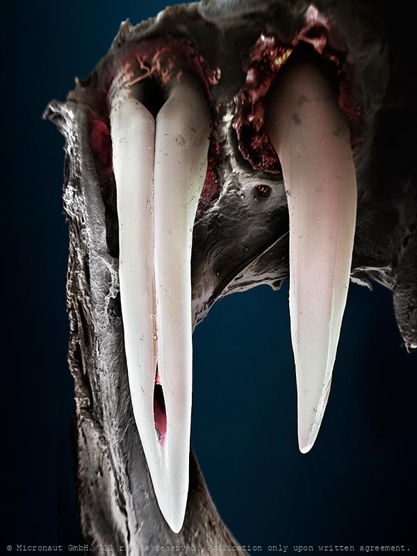
Venemous Bite!
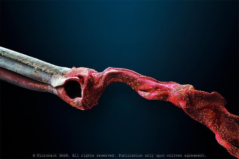
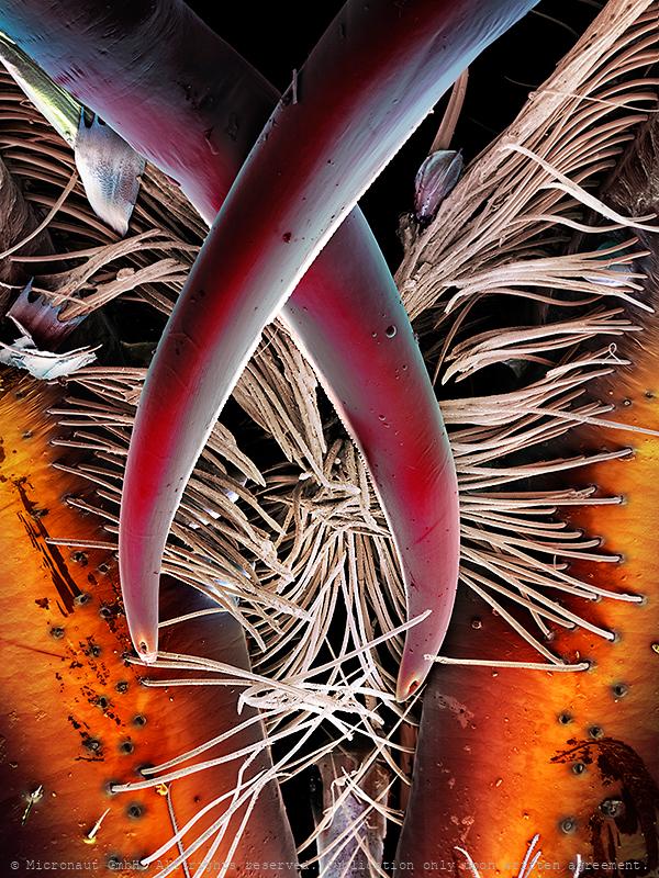
This enlarged microcraph of a Cobra bite is based on Scanning-Electron-Microscopy, and was created and manually colored by Martin Oeggerli (Micronaut). His realistic paintings shed light on invisibly small matters. By dramatically enlarging and exposing his unfamiliar subjects, Martin Oeggerli creates a surrealistic tactility of structures we can neither see, nor touch. Cobra injection: While most snakes are not hazardous to humans, several lineages have evolved venom which is typically delivered by specialized teeth called fangs located on the maxilla (upper jaws). Proteroglyphous snakes (including Cobras) have shortened maxillae bearing few teeth except for a substantially enlarged fang pointing downwards and completely folded around the venom channel, forming a hollow "forward grooved" needle. Because the fangs are only a fraction of an inch long in even the largest species these snakes must hang on, at least momentarily, as they inject their venom. Some spitting cobras have even more modified fangs allowing them to spray venom at an attacker's eyes.
Ant Stinger
Hand-colored scanning electron micrograph of an ant stinger by Martin Oeggerli (Micronaut). Most ants spray or inject a venom. While the Ur-Ant possessed a waspy sting, to paralyze prey before carrying it back to the nest, only rare examples of ants remain with a functional stinger. It is not known for certain, why so many modern species have lost the stinger over time. But a change of the lifestyle of modern ants, moving away from solitary hunting and becoming more dependent on plant food, could be the main reason for the reduction: as the importance of the fierce weapon gradually diminished, it became a burden and was eventually reduced. Examples of modern ants with the functional stinger include Solenopsis (fire ants), Pachycondyla, Myrmecia (bulldog ants), and Paraponera (bullet ants). Some of these are primarily using the stinger as a weapon against vertebrates, and especially in combat with other ant species, the stinger may be used to spray, to wave volatiles about, or to smear a toxin slurry onto enemies, rather than to sting. Stinging ants cause a cutaneous condition that is different from that caused by biting venomous ants. In the case of fire ants, the venom consists of alkaloid and protein components. Their sting is particularly painful, although the bullet ant's sting is considered by some to be the most painful insect sting. In the case of fire ants, the venom consists of alkaloid and protein components, whereas formic acid is only produced within the family Formicidae. Severe allergic reactions can be caused by ant stings in particular and venomous stings in general, including severe chest pain, nausea, severe sweating, loss of breath, serious swelling, fever, dizziness, and slurred speech. They can be fatal if not treated.
The biological gun
Ant stinger (Paltotyreus tarsatus), hand-colored scanning-electron-micrograph by Martin Oeggerli. The stink-ant has volatile material in the venom gland, which includes bitter-tasting cyclic dipeptides (Morgan et al., 2003). Stings are used to forage and for self-defense. Sting pheromones are set free to alarm co-workers upon a sting. Forage workers are also spraying chemical trails in order to recruit co-workers. In general, the stinger is believed to be a rudiment in ants, which have evolved from early solitary hunting hymenopterans. Modern species with a conserved stinger belong to different sub-families, while their closest relatives may have reduced this ancient weapon (especially the more herbivorous species). The bullet-ant (Paraponera clavata) has the most painful sting among insects. According to the Schmidt sting pain index, it is given a "4+" rating, above the tarantula hawk wasp, and according to some victims, equal to being shot, hence the name of the insect. It is described as causing "waves of burning, throbbing, all-consuming pain that continues unabated for up to 24 hours". Poneratoxin, a paralyzing neurotoxic peptide isolated from the venom, affects voltage-dependent sodium ion channels and blocks the synaptic transmission in the central nervous system. It is being investigated for possible medical applications. The Sateré-Mawé people of Brazil use intentional bullet ant stings as part of their initiation rites to become warriors. The ants are first rendered unconscious by submerging them in a natural sedative, and then hundreds of them are woven into gloves made of leaves (which resembles a large oven mitt), stingers facing inward. When the ants regain consciousness, a boy slips the gloves onto his hands. The goal of this initiation rite is to keep the glove on for a full 5 minutes. When finished, the boy's hand and part of his arm are temporarily paralyzed because of the ant venom, and he may shake uncontrollably for days.

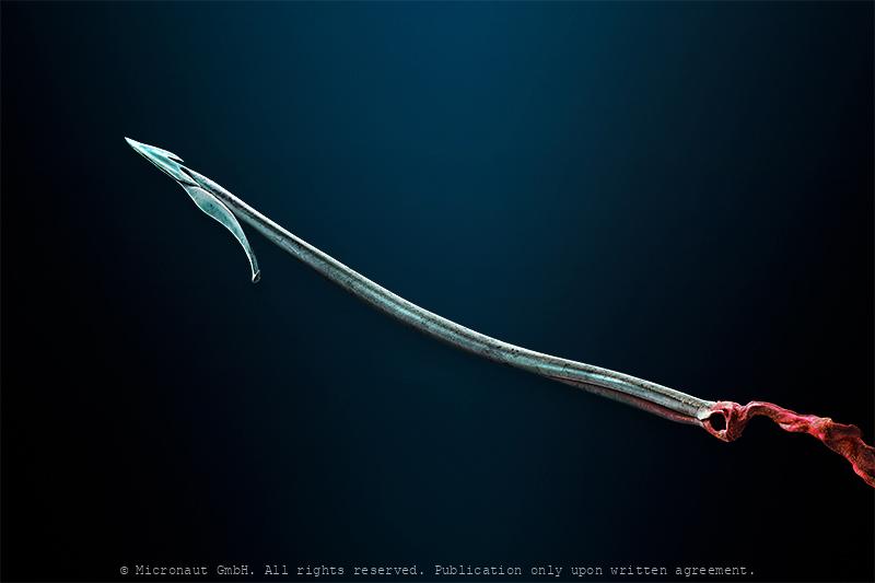
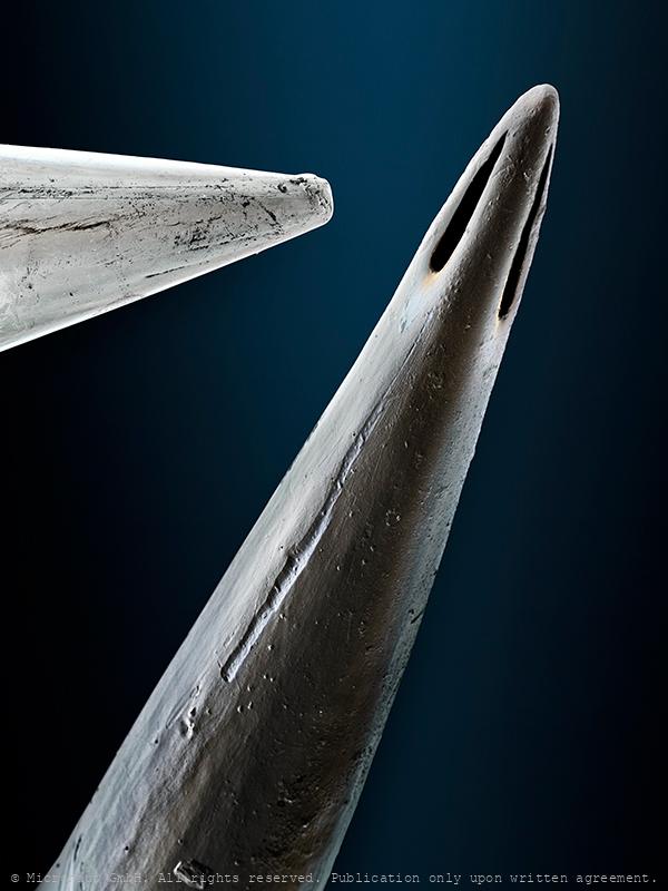
Tip of a hypodermic needle
Tip of a thin surgeon Needle (25g; 1.5; NEO)
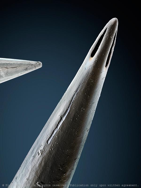
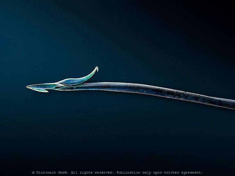
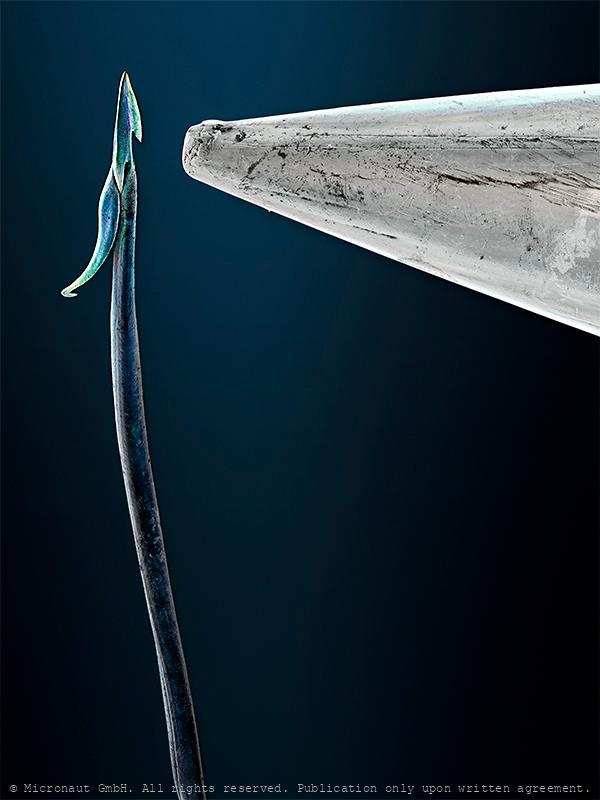
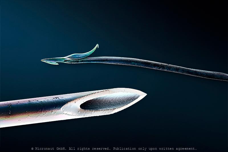
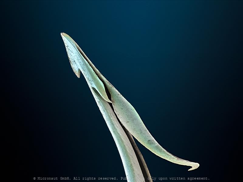
Scorpion injection needle (L. quinquestriatus)
This image shows the tip of a venemous scorpion stinger under the electron microscope. Compared to the sharp Scorpion stinger, a medical injection needle is at best a heavy concrete tunnel tube. Scorpions have eight legs and are easily recognised by the pair of grasping pedipalps and the narrow, segmented tail, often carried in a characteristic forward loop over the back. The tip is connected with a venom gland and injects venom through a pair of elongated holes. Scorpion venoms are neurotoxic cocktails of numerous individual toxins, which attack different targets within a victim’s body: the animals inject relatively large amounts of pain-inducing chemicals, as well as parazyzing- and pre-oral digestion agents, to ultimately kill or paralyze their prey. Scorpion stingers also equipped with mechano-sensitive receptor hairs to detect fine touches, or movements.
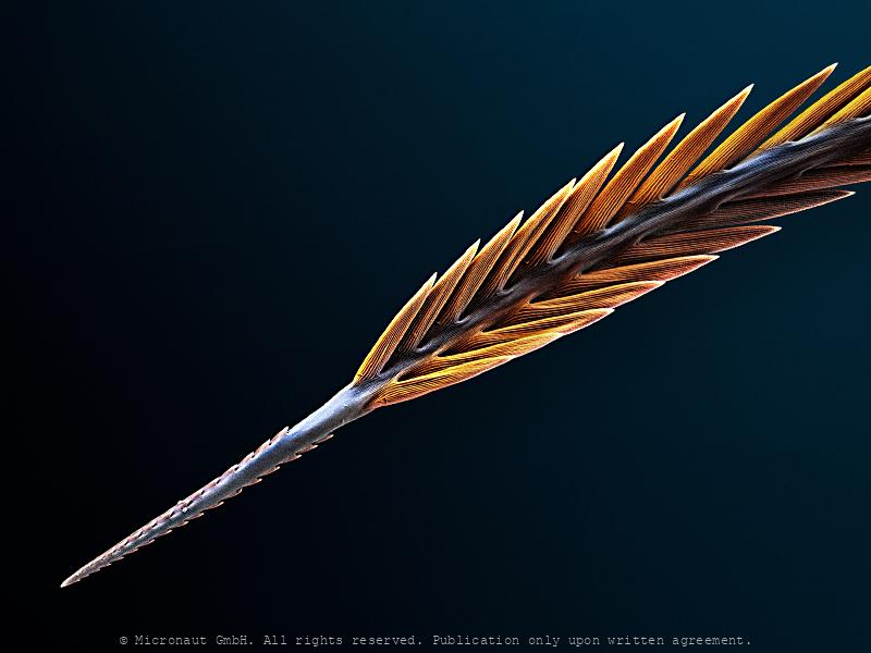
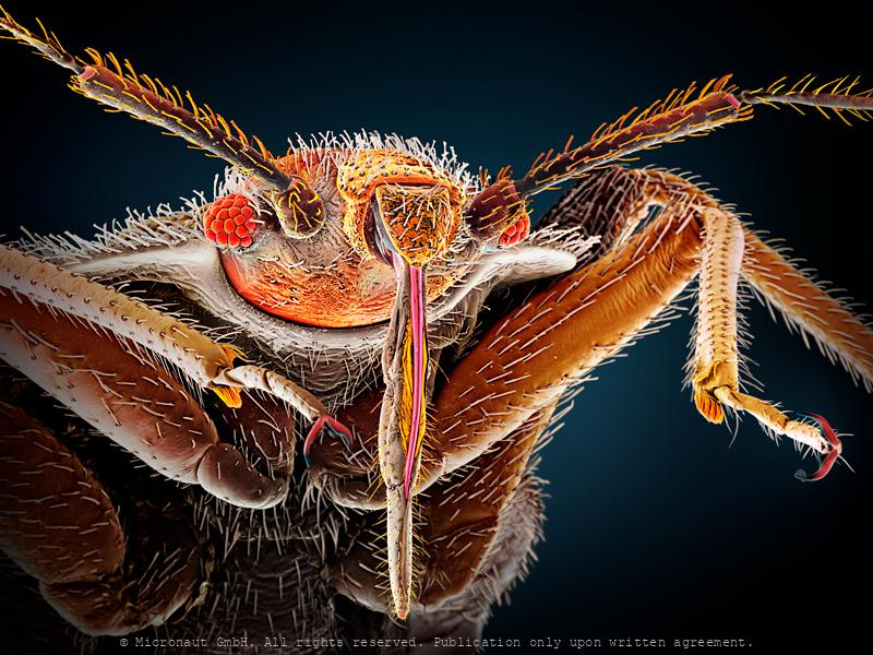
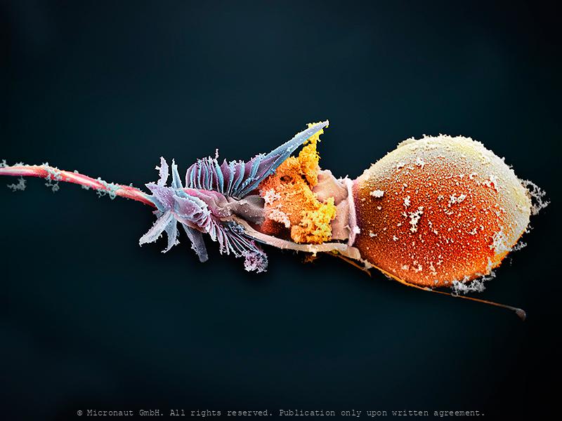
Microarchitecture of the tsetse fly proboscis (Nr.2)
Microarchitecture of the tsetse fly proboscis (Nr.2), manually colored SEM picture by Martin Oeggerli, 2021. Tsetse flies (genus Glossina) are large blood-sucking dipteran flies that are important as vectors of human and animal trypanosomiasis in Africa. The proboscis serves as the developmental site for the infective metacyclic stages of several species of pathogenic trypanosomes that are inoculated into the host with fly saliva. To understand the physical environment in which these trypanosomes develop, you have to look at the microarchitecture of the tsetse proboscis (see image Nr.1, not shown here) and especially at the tip which is usually concealed by surrounding mouthparts (image Nr.2, this is shown here!). SEM technology reveals what's hidden by sheer size - and it turns out the tsetse proboscis isn't just one tiny spear! Instead, the injection tool offers an enormous degree of complexity: a structure called labellum is hidden inside the proboscis and equipped with two 'flippers' at the tip. Sensory bristles and rod like structures are located on the surface of these flippers. They most likely sense tactile information during the sting, and may also register other (chemical) information. A barbed structure (silver) similar to a scarf is located at the very end of the labellum. It contains thousands of tiny hooks. Perhaps the scarf unfolds during the injection and stabilizes the proboscis with its hooks? Or it controls the flow of saliva? Or avoids influx of blood and microorganisms into the hypopharinx? Or it has several functions at once! The more you look at the details and enormous level of sophistication, the more fascinating it becomes (...). Overall, both SEM images confirm that the tsetse proboscis is a formidably armed weapon, specifically adapted for piercing skin, and highlight the utter importance this injection tool has for the fly.
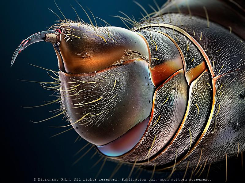
Honey Bee Stinger (Lancet)
This image was produced using scanning electron microscopy in combination with selectively assigning different colors to different structures. By creating a 3D-like photorealistic appearence, the artist aims to expose the delicate fine-structure at the tip of a honey bee (Apis mellifera) stinger. The stinger of a bee is a modified chitinous ovipositor and consists of three parts: a stylus and two barbed slides (or backwards sloping barbed 'lancets'), one on either side of the stylus. In fact, the bee does not have to push the sting in since it is drawn in by the barbed slides which move alternately up and down the stylus so when the barb of one slide has caught and retracts, it pulls the stylus and the other barbed slide into the wound. When the other barb has caught, it also retracts up the stylus pulling the sting further in. This process is repeated until the sting is fully in and even continues after the sting and its mechanism is detached from the bee's abdomen. A sting from a bee (honey bee, bumblebee, sweat bee, etc) can be quite painful. The injected venom or toxin (apitoxin, melittin, histamine, other biogenic amines ) of insects is quite different. Therefore, the body's reaction to a bee sting may differ significantly from one species to another. For about 2 percent of people, a hypersensitivity can develop after being stung, creating a more severe reaction when stung again later. In people with insect sting allergy, a bee sting may trigger a dangerous anaphylactic reaction that is potentially deadly. The sting of a honey bee also releases pheromones that prompt other nearby bees to attack. This alarm process is accelerated if the bee is fatally injured. Although widely believed, a worker honey bee can sting not only once; this partial misconception only happens if the skin of the victim is sufficiently thick and elastic, such as a mammal's. Wasps have stingers with smooth barbs, enabling them to retract the stinger and to sting multiple times. Queen
The Superparasite - Mite (Cheyletidae, Leach 1815)
Title: The Superparasite (Cheyletid sp.) / Der Superparasit (Cheyletid sp.) Beschreibung: Als Superparasit bezeichnet man eine Art (in diesem Fall eine Milbe), welche sich von Parasiten (in diesem Fall andere Milbenarten) ernährt und diese lästigen Biester beseitigt - also aus der Sicht des Wirtes (in diesem Fall ein Specht) ein höchst willkommener Gast. Der Superparasit verfügt über eine Reihe von hochspezialisierten Mundwerkzeugen - vergleichbar mit einem Schweizer Taschenmesser, um seine Beute zu packen, zu immobilisieren, zu verspeisen, oder um sich zu putzen, zu paaren, etc. Dieses Werk stammt aus einer Serie von Milben-Portraits, welche die ausserordentliche Vielfalt dieser Tiergruppe aufzeigt und an den ‘International Photography Awards’ 2011 mit dem 1. Preis im Bereich Spezial-Fotografie ausgezeichnet wurde. Captions: This looks like the cheyletid from Chrysocolaptes lucidus (a woodpecker) from the Philippines. It is predatory and probably feeds on other mites in the plumage of the bird or in its nest. The eyes are a darker red, but are not shown in this picture. The creature also shows a large white patch in the middle of the body but that’s just the excretory glad filled up with guanine crystals, and is ‘optional’ (as it wouldn’t be there after the mite excretes). The things colored in black are the palpal claws. The structures in orange and yellow are two pairs of highly modified setae. All three pairs of structures are used for sensing and manipulating prey. The bird and its parasite have been found on an expedition in 1972 in the Philippines, and were stored in the deep freezer of a candian museum collection until 2011. Then, this specimen was collected, fixed in alcohol and shipped to Switzerland to be analyzed and illustrated by the author (Martin Oeggerli), utilizing a scanning electron microscope (SEM). This work is part of a the artist’s long-term project, i.e. the series of mite portraits which has become famous through th
Pixie dust with a rub in it (Brachypelma smithii)
Urticating hairs are characteristic for the tarantulas of the New World (i.e. North- and South America). To the naked eye, urticating hairs themselves look like minuscule floating lint or dust. However, under the powerful magnification of the scanning-electron-microscope, it turns out they are actually built like barbed spears - and there are six types! Tarantulas have the ability to bite. But before biting, the spider may signal its intention to attack by rearing up into a "threat posture", and by rapidly kicking-up urticating hairs from its abdomen, using the fourth legs in a rapid motion. The tiny hairs gently float up into the air like pixie dust. Touching and inhaling the bristles must be strictly avoided, because they cause severe irritation. To rodents who inhale them, urticating tarantula hairs can actually be fatal. Also, a tarantula may deliberately spread urticating hairs at the entrance to its burrow. Another use is to protect an egg sac, as the hairs help annoy would-be predators, such as fly larvae. It’s important to note that many species of tarantula that have urticating hairs available to them do actually never use them when handled.
Pixie dust with a rub in it (Brachypelma smithii)
Ever heard of urticating hairs? Tarantulas have the ability to bite, but urticating hairs are the invisible enemy. Many species of New World tarantulas (i.e. North and South American), can rapidly kick-up urticating hairs from their abdomens using their fourth legs in a rapid motion. The tiny hairs gently float up into the air like pixie dust. But pixie dust it is not. Avoid breathing the bristles or touching your eyes if you have any bristles on your fingers, as it can cause irritation. The urticating bristles can actually be fatal to rodents who inhale them. The urticating hairs themselves look like minuscule floating lint or dust to the naked eye. Under a scanning-electron-microscope they look like barbed spears, and there are six types. If allowed to land on your skin, they can sometimes cause redness and irritation, although I’ve personally only experienced this with Theraphosa tarantulas. It’s more annoying than anything, but a defense mechanism to avoid if possible. Some species have extremely mild urticating hairs, imperceptible to humans. Others, like the Goliath bird-eating tarantula (Theraphosa sp.) have urticating hairs that can itch and result in a mild rash. Hydrocortisone usually does the trick to alleviate any itching. Also, a tarantula may deliberately spread urticating hairs at the entrance to its burrow. Another use is to protect an egg sac, as the hairs help annoy would-be predators, such as fly larvae. It’s important to note that many species of tarantula that have urticating hairs available to them do actually never use them when handled. This includes Rose hairs and Redknees.
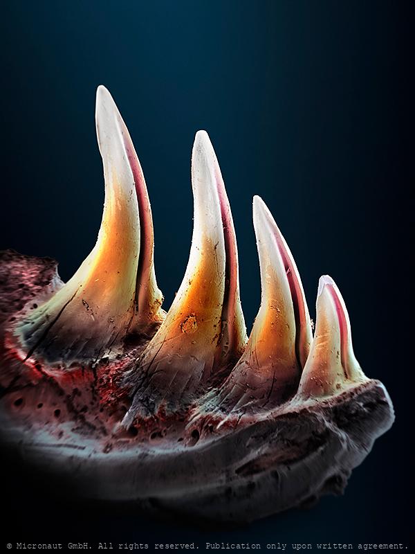
Lizard with Toxic Breath - Gila Monster (Heloderma suspectum)
3D-realistic lower jaw with venemous teeth of the Gila lizard (Heloderma suspectum). This highly enlarged microcraph is based on Scanning-Electron-Microscopy, and was created and manually colored by Martin Oeggerli (Micronaut). His realistic paintings shed light on invisibly small matters. By dramatically enlarging and exposing his tiny and unfamiliar subjects, Martin Oeggerli creates a surrealistic tactility of subjects we can neither see, nor touch. In the Old West, the pioneers believed a number of myths about the Gila monster, including that the lizard had foul or toxic breath and that its bite was fatal. The venomous compounds of the Gila monster are potentially fatal. The lizward deliver their venom by a firm bite and chewing movements with their jaws into the wound. The third to the sixth pair of dentaries (counting from the back) of the lower jaw are equipped with deep grooves and specially shaped to assist in the penetration and increased delivery of venom. The grooves are sustained with venom through small drain ducts, which exit next to the grooved teeth. The venom produces considerable pain and metabolic disorders (blood pressure failure). The potency of the Heloderma venom is believed to be similar to the venom of the Western Diamond Rattlesnake (Crotalus atrox). Exenatide (Exendin-4) is a bioactive peptide identified in the Gila monster venom, that is manufactured now synthetically in large quantities. It serves the treatment of diabetes-type-II (adult-onset diabetes). Type-II patients have difficulty to adjust to physiological glucose levels. Exenatide acts with a different mechanism on the insulin output of the pancreas. This makes type-II patients accessible to therapy. For more information about the biochemistry of Heloderman venom read D. Mebs (Gifttiere 2000; Wissenschaftliche Verlagsges. 2. Auflage, neu bearb. 248-250).
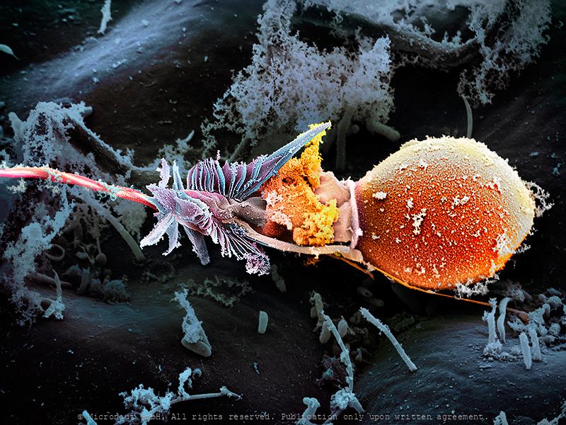
The Magic Bullet - Cnidocyte
The name Cnidaria comes from the Greek word "cnidos", which means stinging nettle. Invisibly small, the cnidocyst which are responsible for the explosive pain caused by jellyfish, remains a mysterious weapon until today. Cnidarians are a varied group of animals including corals, sea anemones, sea pens, sea pansies, jellyfishes, and hydroids. They range among the most beautiful of all marine invertebrates. Many species have a flower-like look and are often considered delicate and soft. But they possess a well-concealed yet potent arsenal of harpoon-like stinging cells. Stings from several species can be very painful, and in some cases, even fatal. Box jellyfish and sea wasps represent the biggest “stinging” threat to divers and swimmers. Portuguese men-of-war are colonial hydroids and their tentacles laced with potent stinging cells are hidden underneath the purple-to-blue float bobbing on the surface. In some specimens the tentacles can reach 20 feet. The virulent stinging cells are easily capable of paralyzing any pelagic fish and in humans can cause extreme pain requiring medical attention. But how does the toxic weapon look like? Cnidocytes are small compartments that house a mini needle-like stinger. When an outside force triggers the stinger, the cell opens, letting ocean water rush in. This causes the stinger to shoot out into what triggered the action; once it’s there, venom is released. All of this happens within a millionth of a second. This picture shows a discharged toxic cnidocyte (i.e. one that has been fired) of a freshwater polyp. It was created with the scanning-electron-microscope by science photographer Martin Oeggerli. Based on the original SEM scan, he colored the picture layer-by-layer, to uncover the delicate structures and shed light on the anatomy of this miniature injection tool.
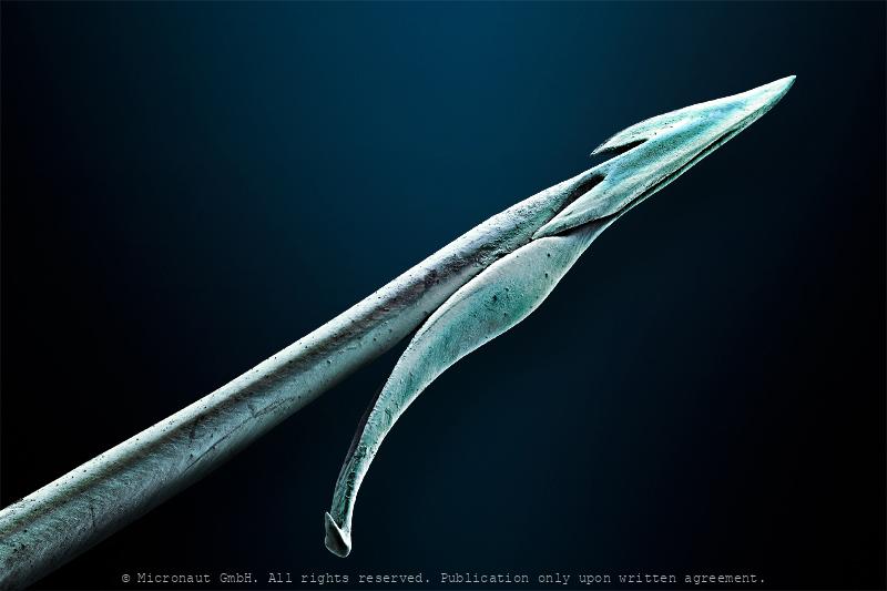
Ambush Weapon - The Conus Snail Radula Tooth (Conus striatus)
This highly enlarged microcraph of a Cone snail harpoon is based on Scanning-Electron-Microscopy, and was created and manually colored by Martin Oeggerli (Micronaut). His realistic paintings shed light on invisibly small matters. By dramatically enlarging and exposing his tiny and unfamiliar subjects, Martin Oeggerli creates a surrealistic tactility of subjects we can neither see, nor touch. Cone snails are marine species that have shells shaped like geometric cones, often with colorful patterning on the surface. All cone snails are venomous. The most dangerous species are the larger cones, which prey on small bottom-dwelling fish, while smaller species prefer to hunt marine worms. Cone snails haunt using a hypodermic needle-like modified radula tooth to attack and paralyze their prey before engulfing it. The tooth, or harpoon, is primarily made of chitin and barbed, similar to a harpoon. The snail can extend its harpoon(s) some distance out from the proboscis when it detects a prey animal nearby. At the same time the harpoon will be loaded with venom from the venom bulb and, still attached to the radula, it can be fired from the proboscis into the prey by a powerful muscular contraction. The venom paralyzes small fish almost instantly. The snail can then retract the radula, drawing the subdued prey into the mouth. Cone snail venoms are mainly peptides, containing hundreds of compounds with different and specific (!) effects. The exact composition of the venom varies widely between species. Cone snail venoms have been propelled into the focus of scientific research as a source of new, medically important substances. The appeal for creating pharmaceutical drugs is the precision and speed with which the various components act; many of the compounds target a particular class of receptor, to the exclusion of any other. This means that, in isolation, they can reliably and quickly produce a particular effect on the body's systems without side effects. How the substance
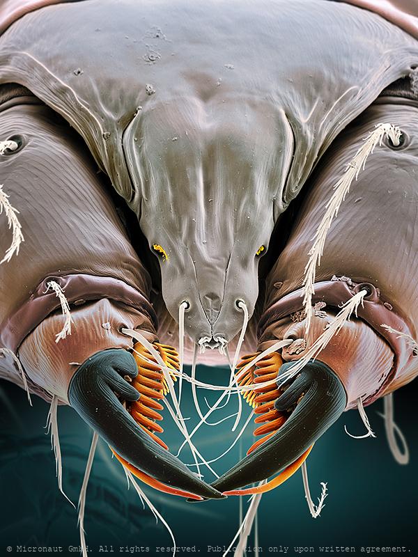
Catflea (Ctenocephalides felis), Nr.2
Frontansicht eines Katzenflohs. Parasiten sind normalerweise auf einen bestimmten Wirt spezialisiert. Katzenflöhe auf Katzen, nur ausnahmsweise werden auch Menschen gestochen (wenn keine Katze in der nähe ist). Portrait of a cat flea (magn. 158:1 if printed 12.5cm high). The cat flea's primary host is the domestic cat, but this is also the primary flea infesting dogs in most of the world. Cat fleas can transmit other parasites and infections to dogs and cats and also to humans. The most prominent of these are Bartonella, murine typhus, and apedermatitis. The tapeworm Dipylidium caninum can be transmitted when a flea is swallowed by pets or humans. In addition, cat fleas have been found to carry Borrelia burgdorferi, the etiologic agent of Lyme disease, but their ability to transmit the disease is unclear.
Bat flea (ev. Ischnopsyllus sp.)
Coloured scanning electron micrograph (SEM) of a Bat flea, showing its head, mandibles and powerful front legs. This parasite was found on a Daubenton's bat (Myotis daubentonii). The mandibles are used to pierce the host's skin and suck blood. As a result of living on nocturnal hosts Bat fleas have no eyes. Interestingly, Bat fleas have relatively short proboscis, measuring less than 200 µm. This is 33% less compared to the Cat flea (Ctenocephalides felis), 50% less compared to the common Rat flea (Xenopsylla cheopis), and even 90% less compared to the common European Mosquito (Culex pipiens). It remains to be determined if insects that parasitize bats evolved a reduced lenght of their blood sucking instruments due to the thin bat wing membranes that are well supplied with blood vessels and lack subcutaneous fat storage or dense fur. For the author the tiny animal reveals the grace of a flamenco dancer and provides a text-book example for the fascination of invisibly small creatures - and especially for those with a notoriously bad reputation generated in childhood from hearsay and fairy tales. Für den Autor hatte der Floh bereits auf der schwarz-weiss Aufnahme die Grazie einer Flamenco-Tänzerin ausgestrahlt. Nun erkennt man dank den Farben noch einiges mehr und ist gleichzeitig erstaunt wie perfekt die einzelnen Strukturen ineinander passen. Augen hat er keine, er lebt meist in völliger Dunkelheit, dafür sind die Fühler und tastenden Mundwerkzeuge prominent entwickelt und das ganze Gesicht ist äusserst fein behaart. Wenn man nichts sieht und auch kein Echolot hat, dann hilft eben der Tastsinn weiter - wir machen es genauso, falls das Licht einmal nicht funktioniert!
Good night, sleep tight, don't let the bed bugs (Cimex lectulari
Bed bug (Cimex lectularius), hand-colored scanning-electron-micrograph image by science artist Martin Oeggerli. Bed bugs are insects from the genus Cimex that feed on blood, usually at night. Their bites can result in a number of health impacts including skin rashes, psychological effects, and allergic symptoms. Bed bug bite symptoms may take between minutes to days to appear and itchiness is generally present. Some individuals may feel tired or have a fever. Typically, uncovered areas of the body are affected. Sensitivity of humans varies from extreme allergic reaction to no reaction at all (about 20%). However, bed bug bites are not known to transmit any infectious disease. Bed bugs have five immature nymph life stages and a final sexually mature adult stage. They are obligatory bloodsuckers and need at least one blood meal in order to advance to the next stage of development. Special mouth parts allow the parasite to saw through the skin and inject saliva with anticoagulants and painkillers. Bed bug secretions can inhibit the growth of some bacteria and fungi, and antibacterial components from the bed bug could be used against human pathogens, and are considered a possible source of pharmacologically active molecules as a resource for the discovery of new drugs.
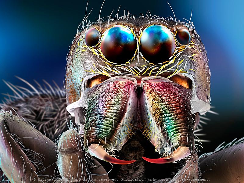
Jumping Spider
Often fuzzy and measuring less than a half inch in body length, Jumping spiders (Salticids) can run, climb, and - jump! Thereby the small predators can achieve stunning distances of more than 50 times its body size. Surprisingly, the extreme jumps are not the result of very strong or muscular legs. Instead, Jumping spiders quickly increase the blood pressure to their legs, which causes the legs to extend and propel the spider through the air. Prior to jumping, whatsoever, the spider glues silk threads to the surface beneath, to allow a quick and safe return to the perch, if required. Interestingly, Salticids rely on an excellent visual system. Like most other spiders, they have eight eyes on their face. An enormous pair in the center facing towards the front, and one adjacent pair of eyes to both sides give them an almost alien appearance. The remaining smaller eyes are located on the dorsal surface of the cephalothorax and allow almost 360° angle panoramic view. In general, animals have developed several different visual systems to accurately and reliably judge distance and depth. Humans, for example, have binocular stereovision. Because our eyes are spaced apart, they receive visual information from different angles, which our brains use to automatically triangulate distances. Other animals, such as insects, adjust the focal length of the lenses in their eyes, or move their heads side to side to create an effect called motion parallax — nearer objects will move across their field of vision more quickly than objects farther away. However, Jumping spiders have eyes that are too close together for binocular stereovision and don’t appear to use motion parallax while hunting. So how are these creatures able to perceive depth? In fact, these amazing creatures have developed a way of gauging distances that appears to be unique in the animal kingdom: rather than having a single layer of photoreceptor cells, as in our case, the spider's retina of the principal e
Spider bite (Chiracanthium punctorium)
Manually colored SEM scan showing the Chelicera of the venemous Yellow-sac spider (Chiracanthium punctorium), by Martin Oeggerli. In Germany, C. punctorium (Dornfingerspinne) is the only "seriously" venomous spider. It is a rare species, only known to occur in notable numbers in the Kaiserstuhl region, the hottest part of the country. C. punctorium can reach a length of about 15 mm, and their painful bite can easily penetrate human skin. However, a bite is not life-threatening, as sometimes spread by the media, and can be compared to a wasp sting. Susceptible persons may have slightly stronger reactions, due to an allergic reaction. Spider venoms are very complex mixtures of diverse chemical compounds ranging from salts to large proteins. In general, spider venom has a high insect toxicity and some antibiotic properties. Therefore, spiders are also regarded a potential source for the development of new insecticides and antibiotics.
Spider Silk, Nr.2
Spider silk is a protein fibre spun by spiders. Spiders use their silk to make webs or other structures, which function as sticky nets to catch other animals, or as nests or cocoons to protect their offspring, or to wrap up prey. They can also use their silk to suspend themselves, to float through the air, or to glide away from predators. Most spiders vary the thickness and stickiness of their silk for different uses. A single spider can produce up to seven different types of silk for different uses. Consisting of mainly protein, silks are about a sixth of the density of steel (1.3 g/cm3). As a result, a strand long enough to circle the Earth would weigh less than 500 grams (18 oz). Making the silk acidic (pH 4) is a protection from fungi and bacteria that would otherwise digest the protein. The spider silk shown here results from combination of four fibres. This hand-painted picture was produced using a scanning electron microscope, by Micronaut (Martin Oeggerli).
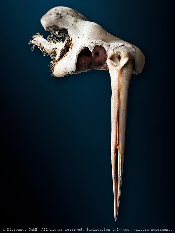
Venemous Bite!
This microcraph of deeply grooved fangs from a venemous snake is based on Scanning-Electron-Microscopy, and was created and manually colored by Martin Oeggerli (Micronaut). His realistic paintings shed light on invisibly small matters. By dramatically enlarging and exposing his unfamiliar subjects, Martin Oeggerli creates a surrealistic tactility of structures we can neither see, nor touch. Boomslangs are native to sub-saharan Africa. They are diurnal and almost exclusively arboreal. Despite Boomslangs are highly venemous, these agile snakes flee from anything too large to eat. Their diet includes chameleons, arboreal lizards, frogs, and occasionally small mammals, birds, and eggs from nesting birds. Many venomous members of the family Colubridae are harmless to humans because of small venom glands and inefficient fangs. However, the boomslang is a notable exception in that it has a highly potent venom, which it delivers through large fangs located in the back of the jaw, equipped with deep grooves to efficientily inject venom. Because boomslang venom is slow-acting, symptoms may not become apparent until many hours after the bite. The maxillary teeth are small anteriorly, seven or eight in number, followed by three very large, grooved fangs situated below each eye. The venom is injected through a deep groove located on the frontside of the large fangs. It is primarily a hemotoxin and disables the coagulation process, causing hemorrhage into tissues such as muslce and brain. The victim may die as a result of internal and external bleeding. Treatment of Boomslang bites involve antivenom, developped during the 40s, and may also require complete blood transfusions, especially after 24 to 48 hours without antivenom. In 1957, the well-known herpetologist Karl Schmidt died after being bitten by a juvenile boomslang which he doubted could produce a fatal dose. Unfortunately, he was wrong. Nevertheless, he made notes on the symptoms he experienced almost to the end.

Honey Bee Stinger (Lancet)
This image was produced using scanning electron microscopy in combination with selectively assigning different colors to different structures. By creating a 3D-like photorealistic appearence, the artist aims to expose the delicate fine-structure at the tip of a honey bee (Apis mellifera) stinger. The stinger of a bee is a modified chitinous ovipositor and consists of three parts: a stylus and two barbed slides (or backwards sloping barbed 'lancets'), one on either side of the stylus. In fact, the bee does not have to push the sting in since it is drawn in by the barbed slides which move alternately up and down the stylus so when the barb of one slide has caught and retracts, it pulls the stylus and the other barbed slide into the wound. When the other barb has caught, it also retracts up the stylus pulling the sting further in. This process is repeated until the sting is fully in and even continues after the sting and its mechanism is detached from the bee's abdomen. A sting from a bee (honey bee, bumblebee, sweat bee, etc) can be quite painful. The injected venom or toxin (apitoxin, melittin, histamine, other biogenic amines ) of insects is quite different. Therefore, the body's reaction to a bee sting may differ significantly from one species to another. For about 2 percent of people, a hypersensitivity can develop after being stung, creating a more severe reaction when stung again later. In people with insect sting allergy, a bee sting may trigger a dangerous anaphylactic reaction that is potentially deadly. The sting of a honey bee also releases pheromones that prompt other nearby bees to attack. This alarm process is accelerated if the bee is fatally injured. Although widely believed, a worker honey bee can sting not only once; this partial misconception only happens if the skin of the victim is sufficiently thick and elastic, such as a mammal's. Wasps have stingers with smooth barbs, enabling them to retract the stinger and to sting multiple times. Queen
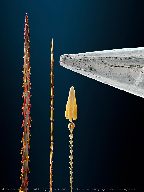
Urticating Hairs
Urticating Hair of the Yellow Tiger Moth Caterpillar (Arctia flava) is shown in the middle, and compared to the tip of a pin (silver, far right). Some fuzzy-looking caterpillars (and other insects) offer a sting by injecting tiny hairs into your skin. Especially caterpillars in the tussock moth subfamily (Lymantriidae), have “urticating hairs” that they use for self-defense. At the tip of each defensive hair is a microscopic barb with a weakened ring at the base, allowing the barb to easily break off in the skin of any animal that grabs onto the caterpillar. These barbs are physically irritating, often causing an itchy-burning sensation, similar to what happens if you touch fiberglass insulation. Some caterpillars take their urticating hairs a step further by connecting them to poison sacs. When the barb breaks off, it also delivers a venom, like the stings of other insects. Seen with a microscope, urticating hair barbs look like miniature hypodermic needles. No wonder picking up a hairy caterpillar can be an uncomfortable experience! This highly enlarged microcraph is based on Scanning-Electron-Microscopy, and was created and manually colored by Martin Oeggerli (Micronaut). His realistic paintings shed light on invisibly small matters. By dramatically enlarging and exposing his tiny and unfamiliar subjects, Martin Oeggerli creates a surrealistic tactility of subjects we can neither see, nor touch.
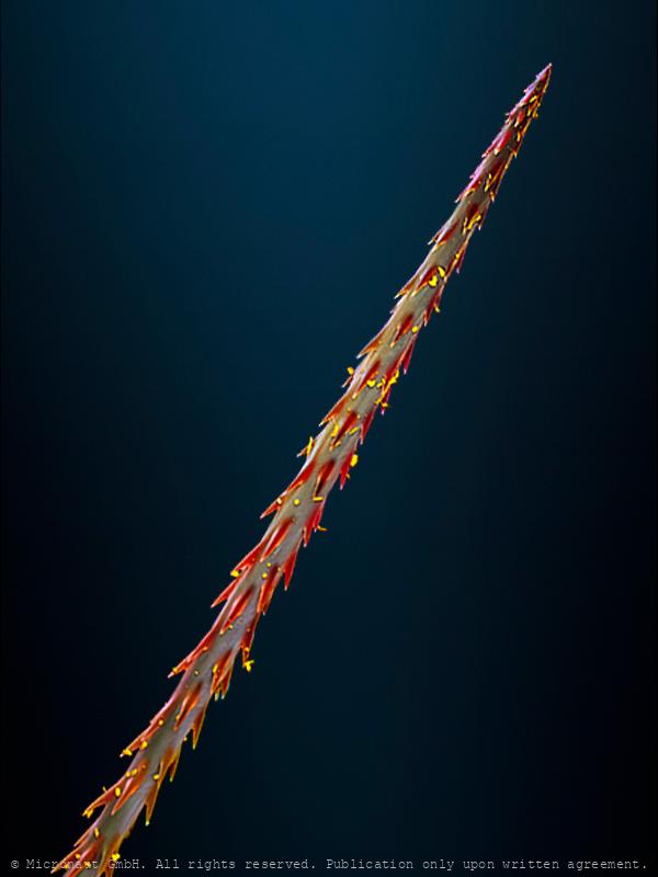
Spike - Urticating hair (unknown species)
Spike - an urticating hair of an unknown species. Found in a garden in Germany. Some fuzzy-looking caterpillars (and other insects) offer a sting by injecting tiny hairs into your skin. Especially caterpillars in the tussock moth subfamily (Lymantriidae), have “urticating hairs” that they use for self-defense. The defensive hair is attached to a weakened ring at the base, allowing the hair to easily break off in the skin of any animal that grabs onto the caterpillar. Barbs are physically irritating, often causing an itchy-burning sensation, similar to what happens if you touch fiberglass insulation. Some caterpillars take their urticating hairs a step further by connecting them to poison sacs. When the barb breaks off, it also delivers a venom, like the stings of other insects. Seen with a microscope, urticating hair barbs look like miniature hypodermic needles. No wonder picking up a hairy caterpillar can be an uncomfortable experience! This highly enlarged microcraph is based on Scanning-Electron-Microscopy, and was created and manually colored by Martin Oeggerli (Micronaut). His realistic paintings shed light on invisibly small matters. By dramatically enlarging and exposing his tiny and unfamiliar subjects, Martin Oeggerli creates a surrealistic tactility of subjects we can neither see, nor touch.
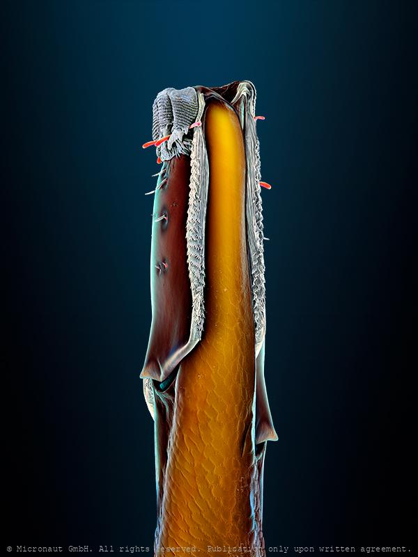
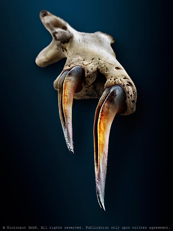
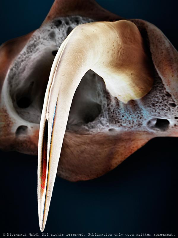
Venemous Bite!
This enlarged microcraph of a Horned viper fang is based on Scanning-Electron-Microscopy, and was created and manually colored by Martin Oeggerli (Micronaut). His realistic paintings shed light on invisibly small matters. By dramatically enlarging and exposing his unfamiliar subjects, Martin Oeggerli creates a surrealistic tactility of structures we can neither see, nor touch. This is likely the most dangerous snake to be found in Europe, due to its large size, long fangs (up to 13 mm) and high venom toxicity. Humans respond rapidly to this venom, as do mice and birds. Lizards are less affected. However, this species is generally not aggressive, and tends not to bite without considerable provocation. Like all pit vipers, the Horned viper is a solenoglyph snake, equipped with a pair of tubular erectile fangs. In solenoglyph snakes, the venom is delivered through a channel located inside the tooth. The venom can be quite toxic, and has both proteolytic and neurotoxic components, containing hemotoxins with blood coagulant properties, similar to and as powerful as in crotaline venom. Other properties include anticoagulant effects, hemoconcentration and hemorrhage. Bites promote symptoms typical of viperid envenomation, such as pain, swelling and discoloration, all of which may be immediate. There are also reports of dizziness and tingling. V. ammodytes venom is used in the production of antivenin for the bite of other European vipers and the snake is farmed for this purpose.
Venemous Bite!
This enlarged microcraph of a pit viper bite is based on Scanning-Electron-Microscopy, and was created and manually colored by Martin Oeggerli (Micronaut). His realistic paintings shed light on invisibly small matters. By dramatically enlarging and exposing his unfamiliar subjects, Martin Oeggerli creates a surrealistic tactility of structures we can neither see, nor touch. Although Copperheads often figure prominently in local scare stories they are shy and nonaggressive in reality. When encountered in the wild, Copperheads lie often motionless in a "pancake coil". When lying on a forest floor among dead leaf litter, their superb camouflage makes them incredibly difficult to spot. Like all pit vipers, the Copperhead is a solenoglyph snake, equipped with a pair of tubular erectile fangs. In solenoglyph snakes, the venom is delivered through a channel located inside the tooth. The venom of the Copperhead is not considered particularly potent (lethal dose of around 100mg). Adult snakes only produce around 70mg, therefore a Copperhead bite is not normally life-threatening, and in most instances of Copperhead bites no antivenin is administered. Instead, the patient is given pain medication and supportive care. Nevertheless, the venom of this serpent causes severe pain and swelling for over a week, as well as localized tissue destruction. Secondary infections are also possible, and if the person is allergic to the venom, an anaphylactic shock can become a life-threatening risk factor.
The Magic Bullet - Cnidocyte
The name Cnidaria comes from the Greek word "cnidos", which means stinging nettle. Invisibly small, the cnidocyst which are responsible for the explosive pain caused by jellyfish, remains a mysterious weapon until today. Cnidarians are a varied group of animals including corals, sea anemones, sea pens, sea pansies, jellyfishes, and hydroids. They range among the most beautiful of all marine invertebrates. Many species have a flower-like look and are often considered delicate and soft. But they possess a well-concealed yet potent arsenal of harpoon-like stinging cells. Stings from several species can be very painful, and in some cases, even fatal. Box jellyfish and sea wasps represent the biggest “stinging” threat to divers and swimmers. Portuguese men-of-war are colonial hydroids and their tentacles laced with potent stinging cells are hidden underneath the purple-to-blue float bobbing on the surface. In some specimens the tentacles can reach 20 feet. The virulent stinging cells are easily capable of paralyzing any pelagic fish and in humans can cause extreme pain requiring medical attention. But how does the toxic weapon look like? Cnidocytes are small compartments that house a mini needle-like stinger. When an outside force triggers the stinger, the cell opens, letting ocean water rush in. This causes the stinger to shoot out into what triggered the action; once it’s there, venom is released. All of this happens within a millionth of a second. This picture shows a discharged toxic cnidocyte (i.e. one that has been fired) of a freshwater polyp. It was created with the scanning-electron-microscope by science photographer Martin Oeggerli. Based on the original SEM scan, he colored the picture layer-by-layer, to uncover the delicate structures and shed light on the anatomy of this miniature injection tool.
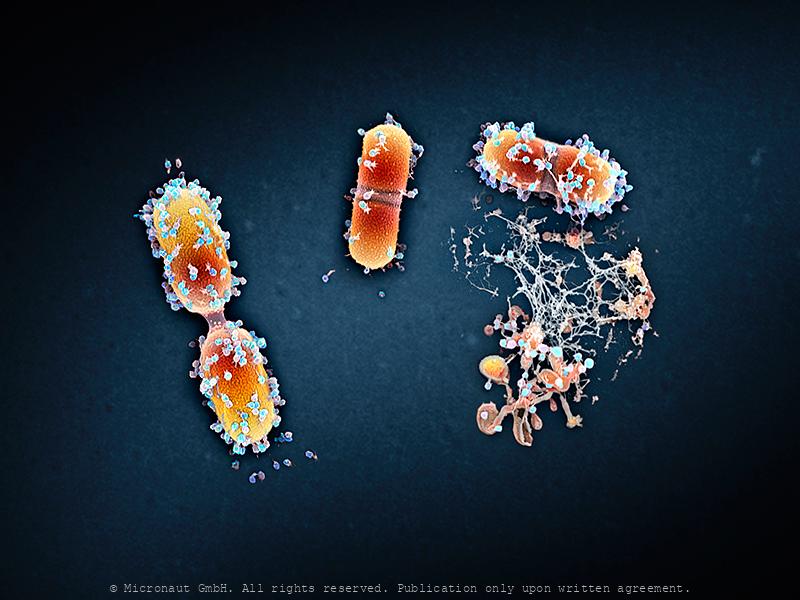
Viral infection - The Parasites of Parasites of Parasites... Nr.1
A bacteriophage is a virus that infects and replicates within a bacterium. It is composed of proteins that encapsulate a DNA or RNA genome. Bacteriophages ('phages') have relatively simple structures and their genomes encodes something between four and several hundred genes. To replicate, phages inject their genome into the cytoplasm of a bacteria. Bacteriophages are among the most common and diverse entities in the biosphere. They are widely distributed in locations populated by bacterial hosts, such as soil or the intestines of animals. One of the densest natural sources for phages and other viruses is sea water. This hand colored scanning-electron-micrograph shows bacteria which are heavily infected by bacteriophages. At the lower right hand side, a bacterial cell has been broken open (lysed) and destroyed. As soon as the cell is destroyed, the phage progeny can find new hosts to infect.
Viral infection - The Parasites of Parasites of Parasites... Nr.1
A bacteriophage is a virus that infects and replicates within a bacterium. It is composed of proteins that encapsulate a DNA or RNA genome. Bacteriophages ('phages') have relatively simple structures and their genomes encodes something between four and several hundred genes. To replicate, phages inject their genome into the cytoplasm of a bacteria. Bacteriophages are among the most common and diverse entities in the biosphere. They are widely distributed in locations populated by bacterial hosts, such as soil or the intestines of animals. One of the densest natural sources for phages and other viruses is sea water. This hand colored scanning-electron-micrograph shows bacteria which are heavily infected by bacteriophages.
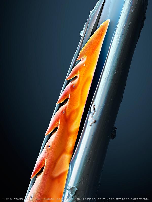
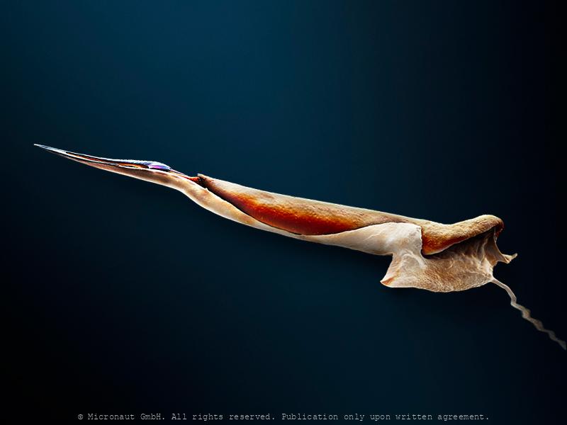
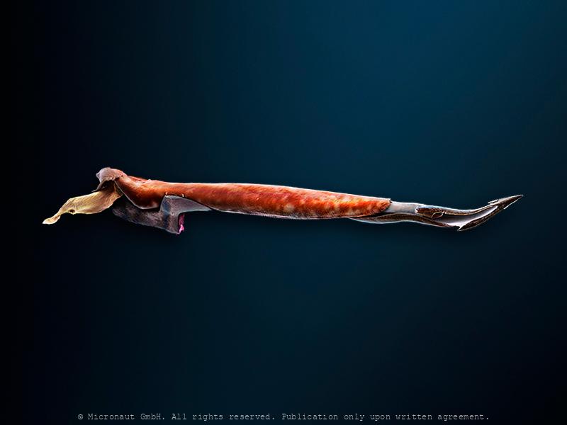
Deadly Harpoon (Conus striatus)
This highly enlarged microcraph of a Cone snail harpoon is based on Scanning-Electron-Microscopy, and was created and manually colored by Martin Oeggerli (Micronaut). His realistic paintings shed light on invisibly small matters. By dramatically enlarging and exposing his tiny and unfamiliar subjects, Martin Oeggerli creates a surrealistic tactility of subjects we can neither see, nor touch. Cone snails are marine species that have shells shaped like geometric cones, often with colorful patterning on the surface. All cone snails are venomous. The most dangerous species are the larger cones, which prey on small bottom-dwelling fish, while smaller species prefer to hunt marine worms. Cone snails haunt using a hypodermic needle-like modified radula tooth to attack and paralyze their prey before engulfing it. The tooth, or harpoon, is primarily made of chitin and barbed, similar to a harpoon. The snail can extend its harpoon(s) some distance out from the proboscis when it detects a prey animal nearby. At the same time the harpoon will be loaded with venom from the venom bulb and, still attached to the radula, it can be fired from the proboscis into the prey by a powerful muscular contraction. The venom paralyzes small fish almost instantly. The snail can then retract the radula, drawing the subdued prey into the mouth. Cone snail venoms are mainly peptides, containing hundreds of compounds with different and specific (!) effects. The exact composition of the venom varies widely between species. Cone snail venoms have been propelled into the focus of scientific research as a source of new, medically important substances. The appeal for creating pharmaceutical drugs is the precision and speed with which the various components act; many of the compounds target a particular class of receptor, to the exclusion of any other. This means that, in isolation, they can reliably and quickly produce a particular effect on the body's systems without side effects.
Ambush Weapon - The Conus Snail Radula Tooth (Conus striatus)
This highly enlarged microcraph of a Cone snail harpoon is based on Scanning-Electron-Microscopy, and was created and manually colored by Martin Oeggerli (Micronaut). His realistic paintings shed light on invisibly small matters. By dramatically enlarging and exposing his tiny and unfamiliar subjects, Martin Oeggerli creates a surrealistic tactility of subjects we can neither see, nor touch. Cone snails are marine species that have shells shaped like geometric cones, often with colorful patterning on the surface. All cone snails are venomous. The most dangerous species are the larger cones, which prey on small bottom-dwelling fish, while smaller species prefer to hunt marine worms. Cone snails haunt using a hypodermic needle-like modified radula tooth to attack and paralyze their prey before engulfing it. The tooth, or harpoon, is primarily made of chitin and barbed, similar to a harpoon. The snail can extend its harpoon(s) some distance out from the proboscis when it detects a prey animal nearby. At the same time the harpoon will be loaded with venom from the venom bulb and, still attached to the radula, it can be fired from the proboscis into the prey by a powerful muscular contraction. The venom paralyzes small fish almost instantly. The snail can then retract the radula, drawing the subdued prey into the mouth. Cone snail venoms are mainly peptides, containing hundreds of compounds with different and specific (!) effects. The exact composition of the venom varies widely between species. Cone snail venoms have been propelled into the focus of scientific research as a source of new, medically important substances. The appeal for creating pharmaceutical drugs is the precision and speed with which the various components act; many of the compounds target a particular class of receptor, to the exclusion of any other. This means that, in isolation, they can reliably and quickly produce a particular effect on the body's systems without side effects.
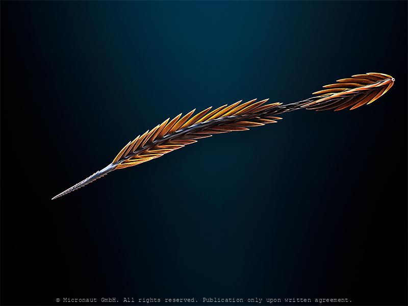
Marine snail harpoon (Profundiconus cf. teramachii)
Venemous radular tooth of a marine Cone snail (Profundiconus cf. teramachii). This highly enlarged microcraph is based on Scanning-Electron-Microscopy and was created and manually colored by Martin Oeggerli (Micronaut). His realistic paintings shed light on invisibly small matters. By dramatically enlarging and exposing his tiny and unfamiliar subjects, Martin Oeggerli creates a surrealistic tactility of subjects we can neither see, nor touch.Cone snails are marine species that have shells shaped like geometric cones, often with colorful patterning on the surface. All cone snails are venomous. The most dangerous species are the larger cones, which prey on small bottom-dwelling fish, while smaller species prefer to hunt marine worms. Cone snails haunt using a hypodermic needle-like modified radula tooth to attack and paralyze their prey before engulfing it. The tooth, or harpoon, is primarily made of chitin and barbed, similar to a harpoon. The diet of Profundiconus is unknown. Presumed vermivorous. However, there are reports of finding the beak of an small octopus in the gut of a specimen of Profundiconus smirnoides. Also, the radular morphology of Pygmaeconus resembles very much that of Californiconus californicus, a species known to be a generalist feeder, preying on worms, molluscs, fishes and even shrimps. However, the relationship between the diet and the radular morphology might not be so straightforward as initially expected.Cone snail venoms are mainly peptides, containing hundreds of compounds with different and specific (!) effects. The exact composition of the venom varies widely between species. Cone snail venoms have been propelled into the focus of scientific research as a source of new, medically important substances. The appeal for creating pharmaceutical drugs is the precision and speed with which the various components act; many of the compounds target a particular class of receptor, to the exclusion of any other. This means that, in isolation, the
Marine snail harpoon (P. neocaledonicus)
Venemous radular tooth of a marine Cone snail (Profundiconus neocaledonicus). This highly enlarged microcraph is based on Scanning-Electron-Microscopy and was created and manually colored by Martin Oeggerli (Micronaut). His realistic paintings shed light on invisibly small matters. By dramatically enlarging and exposing his tiny and unfamiliar subjects, Martin Oeggerli creates a surrealistic tactility of subjects we can neither see, nor touch. Cone snails are marine species that have shells shaped like geometric cones, often with colorful patterning on the surface. All cone snails are venomous. The most dangerous species are the larger cones, which prey on small bottom-dwelling fish, while smaller species prefer to hunt marine worms. Cone snails haunt using a hypodermic needle-like modified radula tooth to attack and paralyze their prey before engulfing it. The tooth, or harpoon, is primarily made of chitin and barbed, similar to a harpoon. The diet of Profundiconus is unknown. Presumed vermivorous. However, there are reports of finding the beak of an small octopus in the gut of a specimen of Profundiconus smirnoides. Also, the radular morphology of Pygmaeconus resembles very much that of Californiconus californicus, a species known to be a generalist feeder, preying on worms, molluscs, fishes and even shrimps. However, the relationship between the diet and the radular morphology might not be so straightforward as initially expected. Cone snail venoms are mainly peptides, containing hundreds of compounds with different and specific (!) effects. The exact composition of the venom varies widely between species. Cone snail venoms have been propelled into the focus of scientific research as a source of new, medically important substances. The appeal for creating pharmaceutical drugs is the precision and speed with which the various components act; many of the compounds target a particular class of receptor, to the exclusion of any other. This means that, in isolatio
Scorpion injection needle (L. quinquestriatus)
This highly enlarged micrograph of a venemous Scorpion stinger is based on Scanning-Electron-Microscopy, and was created and manually colored by Martin Oeggerli (Micronaut). Compared to the sharp stinger, the tip of a needle looks like a blunt instrument at best. Scorpions have eight legs and are easily recognised by the pair of grasping pedipalps and the narrow, segmented tail, often carried in a characteristic forward loop over the back. The tip is connected with a venom gland and injects venom through a pair of elongated holes. Scorpion venoms are neurotoxic cocktails of numerous individual toxins, which attack different targets within a victim’s body: the animals inject relatively large amounts of pain-inducing chemicals, as well as parazyzing- and pre-oral digestion agents, to ultimately kill or paralyze their prey. Scorpion stingers also equipped with mechano-sensitive receptor hairs to detect fine touches, or movements.

Cutting Edge
Forged Steel of a Guarda Knife (cutting edge) hand colored SEM picture by Martin Oeggerli (coloration) and Theo Bühler (SEM scan).

Pin tip
Tip of a metal pin