Dr.Oeggerli’s Beautiful Little Monsters
Some people find Dr. Martin Oeggerli’s monsters a little frightening. I see beauty in them, perfectly shaped by evolution. I also see a hint of Charles Darwin and Albrecht Dürer: the scientist and the artist. Mites don’t have a particularly nice reputation, but how well do we really know them? Oeggerli wants to help us to see past our preconceptions and beyond the limits of our own eyes. With a scanning electron microscope (SEM), he explores the microcosmos—the tiny, unseen world that’s smaller than the period at the end of this sentence. His work (reminds) me of Dürer’s rhinoceros. This strange creature, unknown to Europe in 1515, captured the imagination of the entire continent partly because the artist’s famous etching made it visible. Whatever his work of artistic interpretation lacked in accuracy, it made up for with its sheer beauty, its strangeness, and its power to inspire the imagination. Dürer was an artist drawn toward science, or at least animated by its principal ingredient: curiosity. Oeggerli is a man of science drawn toward artistic interpretation: Art multiplies science for him.
Continue reading, text written by Todd James and published by National Geographic, 22 Jan. 2015.
«Many people have already commented very favourably on your beautiful images in the National Geographic article! I have been proud to tell them that Dave and I were involved in the creation of Mighty Mites by providing specimens and information. Thanks very much.»
Heather Proctor, PhD
Prof. at University of Alberta, Canada
Micronaut images are rights-managed. If you want to get a quote, please contact us, providing the following information: (1) image name, (2) specific use, (3) industry, (4) distribution area, (5) format, (6) circulation or print run, and (7) duration.
Please note that we cannot answer incomplete requests. Thank you.
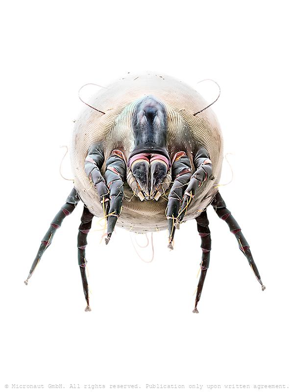
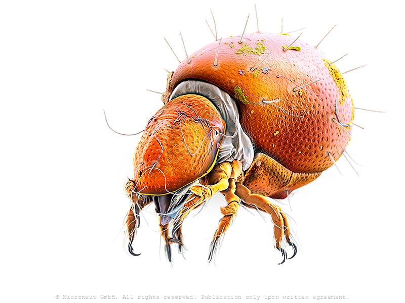
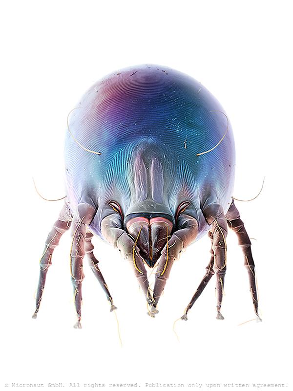
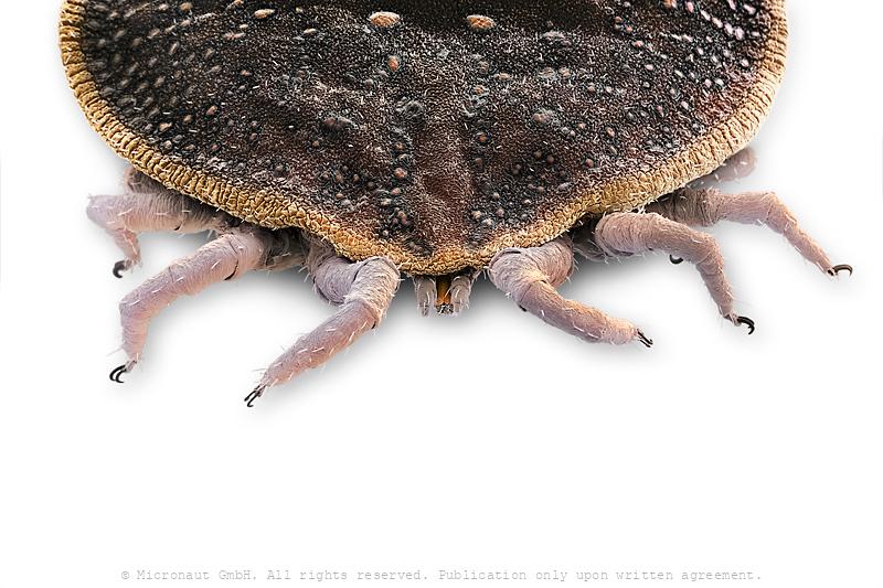
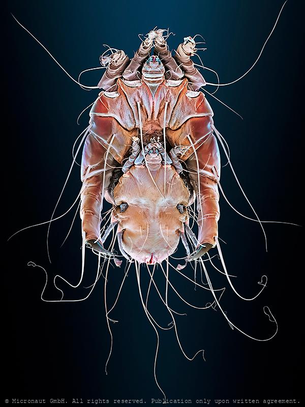
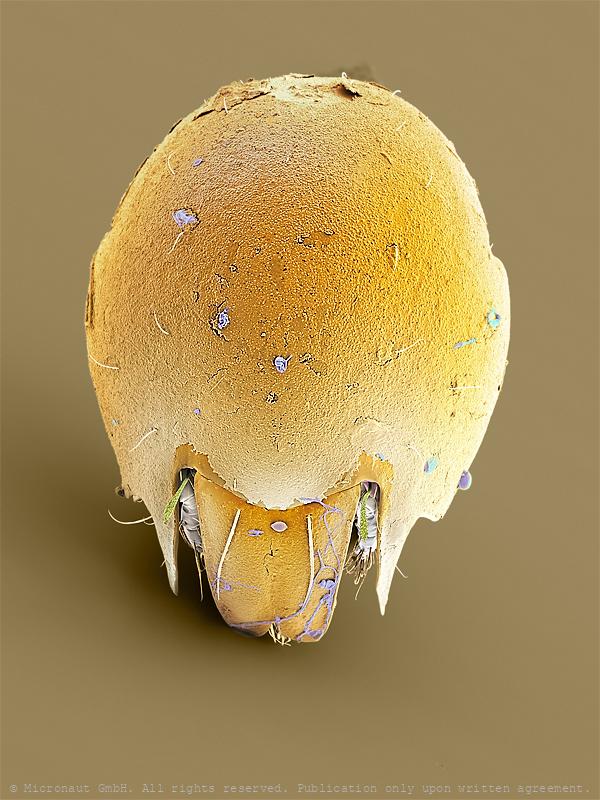
House Dust Mite (D. farinae) Nr.3
Hand-colored scanning electron micrograph of a House Dust Mite (D. farinae) by Martin Oeggerli - The house dust mite is a cosmopolitan pyroglyphid mite that lives in human habitation. Dust mites typically measure between 0.2–0.3 millimetres (0.008–0.012 in) in length and feed on organic detritus, such as flakes of shed human skin, and flourish in the stable environment of dwellings. House dust mites are a common cause of asthma and allergic symptoms worldwide. The mite's gut contains potent digestive enzymes (notably proteases) that persist in their feces and are major inducers of allergic reactions such as wheezing. The mite's exoskeleton can also contribute to allergic reactions. There are two different species which occur widely: the European house dust mite (Dermatophagoides pteronyssinus) and the American house dust mite (Dermatophagoides farinae); a third species (Euroglyphus maynei) also occurs worldwide. Unlike scabies mites or skin follicle mites, house dust mites do not burrow under the skin and are not parasitic. Dieses Bild wurde mittels Raster-Elektronen-Mikroskopie aufgenommen und nachträglich manuell koloriert. Es zeigt eine aufgeblasene drohende Hausstaubmilbe.
Mite (Scutarid)
Scutacarid! These are phoretic on larger arthropods, including other mites, which they ride from place to place in search of fresh fungi and other microbes to eat.
Parazercon sp.
This is a coloured scanning electron micrograph (SEM) of a predatory soil-dwelling mite, Mesostigmata: Zerconidae: possibly a Parazercon sp. Mites are a highly adaptable group of Arthropoda that are related to spiders and scorpions, have eight legs and are usually smaller than a full stop. Despite representing one of the most diverse groups within the animal kingdom (>20'000 species), mites are notoriously overlooked due to the diminutive size. Millions of dust mites live inside furniture and fabric in the average home. The dead bodies and excrement of dust mites can cause allergic reactions to household dust. Other mites are parasites, whereas soil mites form part of the great diversity of organisms that contribute to the break down of plant material. The grey appendages in the central part of the image are the pedipalps. The seta („hairs“) with the modified tip belongs to the tarsus of the first pair of legs. In ticks, this area contains Haller’s organ – a complex of pits and modified setae that sense things like CO2 and infrared. The function of the setal cluster at the tip of tarsus nr.1 is not well studied in Mesostigmata, but undoubtedly contains a variety of chemosensory setae. Sometimes they have a swollen tip like the one here. The yellow, tongue-like process in the area of the chelicera is probably the palpal apotele. This is a remnant of the claw of the palps and is produced between the palpal tibia and tarsus. It is used to groom the chelicerae. In zerconids it is bifurcate and looks a bit like an index and second finger would if held slightly apart. The thin line of grey probably represents the space between both parts of this bifurcate palpal apotele (or „tongue“).
Soil mite (Astigmata), Nr.2
this is an adult female Astigmata of some sort. Without seeing the dorsal setae, it may not be possible to get it to family.

Catflea (Ctenocephalides felis), Nr.2
Frontansicht eines Katzenflohs. Parasiten sind normalerweise auf einen bestimmten Wirt spezialisiert. Katzenflöhe auf Katzen, nur ausnahmsweise werden auch Menschen gestochen (wenn keine Katze in der nähe ist). Portrait of a cat flea (magn. 158:1 if printed 12.5cm high). The cat flea's primary host is the domestic cat, but this is also the primary flea infesting dogs in most of the world. Cat fleas can transmit other parasites and infections to dogs and cats and also to humans. The most prominent of these are Bartonella, murine typhus, and apedermatitis. The tapeworm Dipylidium caninum can be transmitted when a flea is swallowed by pets or humans. In addition, cat fleas have been found to carry Borrelia burgdorferi, the etiologic agent of Lyme disease, but their ability to transmit the disease is unclear.
The First Dsease of Humans (Scabies, 1687)
This is a hand colored scanning electron micrograph (SEM) of Sarcoptes scabiei, the itch mite, which is a parasitic arthropod burrowing into human skin, thereby causing scabies. The discovery of the itch mite in 1687 marked scabies as the first disease of humans with a known cause. Other mammals can also become infected, including dogs, cats, ungulates, wild boars, bovids, wombats, koalas, and great apes. About 2% of the British population is thought to be infected with these mites, which take about 25 minutes to an hour to burrow into the skin. The burrowing is carried out using the mouth parts and special cutting surfaces on the front legs. A burrowing mite anchors with suckers on its feet, furrows and spines, located on its body surface. Eggs are laid in small numbers as the mite burrows. As these hatch, six-legged larvae climb out on to the skin and search for hair follicles, where they feed and develop. In the hair follicles, the larvae show the first nymphal stages, with eight legs.
Soil mite Nr.3 (Eobrachychthonius)
This mite is a Brachychthoniidae and possibly Eobrachychthonius, one of the few oribatids that retain a lateral ocellus that is a simple eye between the seta and bothridium (shown in orange). Eobrachychthonius belongs to the order of the Oribatida, better known as beetle mites or moss mites. Most of them are heavily armored by thick body plates which help to reduce the chance that something eats them. Internally, many are filled with toxins to the same end. Poison dart frogs get their toxins from oribatid mites (and ants), so the defenses don’t always work to their advantage, but often enough for them to have persisted form over 450 million years! Mites secrete an outermost layer of waxes and proteins called the cerotegument (as shown in yellow/green on legs and on remaining body parts). The cerotegument of species found in dry microhabitats has pustulate ornamentation which look a bit like Bucky Balls at high magnification – very similar to some of the types of brochosomes found on Cicadellidae. Likely the cerotegument are water-repellent structures analogous to those found on the upper side of the leaves of water lilies. The 'ear' or 'antenna' is a special seta called a sensillus that can be moved and is sensitive to airborne vibrations. The very deep base (the 'screw-threaded-tube') for the sensillus is called a bothridium. It provides extra support for the sensillus to allow it to vibrate but not to snap off. There is another sensillus and bothridium on the other side of the mite's 'head'. Almost all oribatid species have a pair of sensilli. Unusual for oribatids, Brachychthoniidae can be rather colourful: in the species Eobrachychthonius latior the sclerites are orange in adults and immatures are violet. Suprapleural plates that may be present appear to be infolded, therefore it is not sure if this mite belongs to the genus Eobrachychthonius. The mouthparts on either side of the chelicerae are called pedipalps – homologous to the pincers of sco
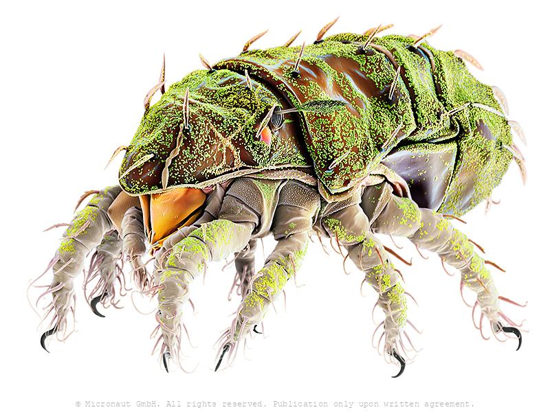
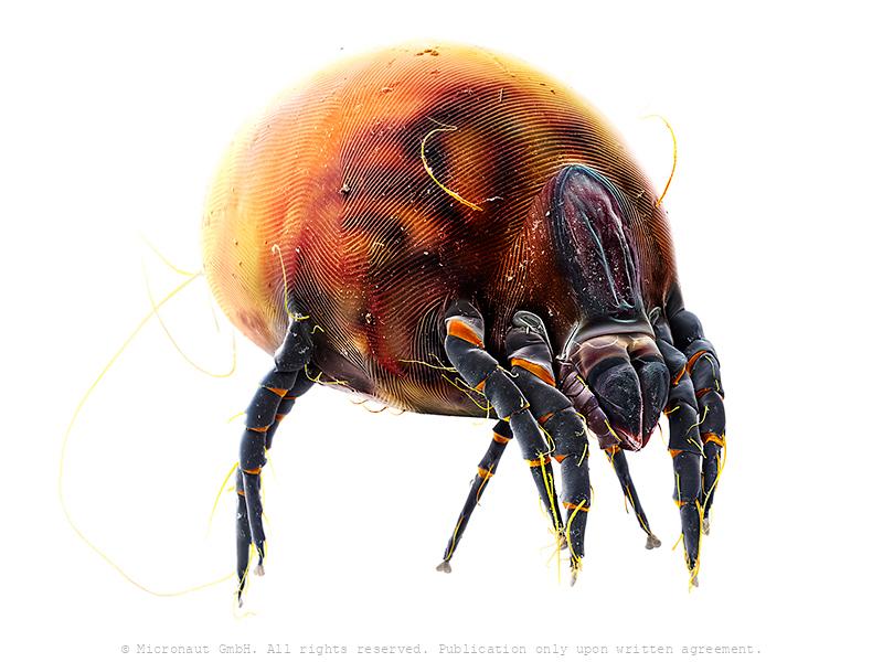
Predatory Mite (Gamasellus sp.), Nr.1
Colored Scanning Electron Micrograph of a predatory mite (Gamasellus sp.). You can be grateful this animal measures only app. 0.18mm in diameter, because it has a horrible bite: adult Gamasellus eat Springtails (Collembola sp.) of the same size! Compared to juveniles, adults focus on larger prey and eat more often (juvenile: 1 Springtail every 12 days, adult: 1 Springtail every 3 days). Gamasellus is an agile predator and attacks quickly through looping his elongated forelegs over the prey. After this, the chelicerae are moved forward to "kiss" the prey, and inject digestion fluid through a pinhole at the tip of the upper left and right chelicerae. The poisonous fluid tranquilizes and eventually kills and digests the prey. Such a procedure is called pre-oral digestion and it is common in the class of Arachnida, especially in spiders. A multitude of mechanosensory setae ("hairs") are located on the forelegs and pedipalps. They serve the blind predator to monitor the exact position of its prey and make sure it is held in position until the deadly cocktail is effectively working.
Watermite (Frontipoda sp.), Nr.2
Water mite from Alberta, Canada. This animal belongs to the Prostigmata: Oxidae: Frontipoda sp. Mites are a highly adaptable group of Arthropoda that are related to spiders and scropions, have eight legs and are usually smaller than a full stop. Despite representing one of the most diverse groups within the animal kingdom (>20’000 species), mites are notoriously overlooked due to the diminutive size. Millions of dust mites live inside furniture and fabric in the average home. The dead bodies and excrement of dust mites can cause allergic reactions to household dust. While some mites are parasites and others form part of the great diversity of organisms that contribute to the break down of plant material, Frontipoda is a predatory mite which lives in small ponds and feeds on larval midges. Interestingly, Frontipoda has two pairs of bright red eyes (each with two lenses, the bottom one larger than the top), which lie below the integument. They are clearly visible in the live animal. This work is part of Micronaut’s ‘Cursed Knights’-series which shows close-up portraits of mites and has become famous at the IPA / Lucy Awards, after winning the 1st prize in the ‘Special Photography-Category’. Furthermore, Micronaut has been awarded ‘International Photographer of the Year’ in 2011, due to these unique works.
Box Mite (Atropacarus sp.), Nr.1
Adults are slow-moving, deliberate, and heavily encumbered in armour. One remarkable type of defence that has evolved at least three times in the Oribatida is called ptychoidy (more Greek, ‘ptych’, a fold). Ptychoid mites are able to fold their legs into their bodies and close the anterior shell-like aspis over the legs, giving rise to a less English-tongue-twisting name, ‘box mites’. Most Boxmite species feature a clean body but some cover themselves with a layer of soil. Presumably this serves as a visual or, more likely, tactile camouflage that increases the chance a predator will move on (‘get along now, nothing but a bit of dirt, your dinner is elsewhere’).
Box Mite (Atropacarus sp.), Nr.2
Adults are slow-moving, deliberate, and heavily encumbered in armour. One remarkable type of defence that has evolved at least three times in the Oribatida is called ptychoidy (more Greek, ‘ptych’, a fold). Ptychoid mites are able to fold their legs into their bodies and close the anterior shell-like aspis over the legs, giving rise to a less English-tongue-twisting name, ‘box mites’. Most Boxmite species feature a clean body but some cover themselves with a layer of soil. Presumably this serves as a visual or, more likely, tactile camouflage that increases the chance a predator will move on (‘get along now, nothing but a bit of dirt, your dinner is elsewhere’).
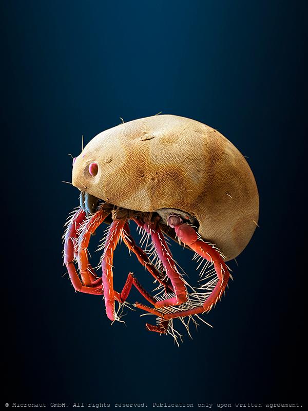
Superparasite (Alleustathia ungulata)
female Alleustathia cf. ungulata (Eustathiidae), a feather mite from the Glossy Swiftlet Collocalia esculenta from the Philippines. The taxonomic illustrations of this species do not show the deep rugosities that are evident on the dorsal terminus of these specimens; however, the male mites that I mounted from this collection do key to A. ungulata. If you want to play it safe, you can call them Alleustathia sp.
Parasitic frisbee (Varroa destructor)
Varroa destructor is an external parasitic mite that attacks the honey bees (Apis cerana and Apis mellifera). The disease caused by the mites is called varroatosis. The species was discovered in Java in 1904 and the man who described the mite named it Varroa jacobsoni, after the collector. Apparently, he could not imagine the enormous threat the mite would become some hundred years later. For a long time, this was of little interest to anybody. Within the species there is little morphologic diversity. However, genetic studies in the late 1990s identified a second species within V. jacobsoni which was much more virulent to Honey bees and was therefore named V. destructor. Varroa destructor reproduces in honey bee colonies. The image shows an adult female which are three times larger than adult males. The parasite attaches to the body of a bee and weakens the host by sucking hemolymph. In this process, RNA viruses such as the deformed wing virus (DWV) spread to bees. A significant mite infestation leads to the death of a honey bee colony. The Varroa mite is the world's most devastating pest of Western honey bees. Varroa mite life cycle has two stages. During the phoretic stage (5-11 days), mites ride on adult workers or drones, at the same time feeding on blood (hemolymph) from bees, usually from the inter-segmental membrane on the abdomen. Mites change hosts (hop from one bee to another) often and this contributes to transmission of various viruses. The other stage is the reproductive stage. The mite invades a worker or drone larvae cell just before the cell being capped. Once inside, it hides in the brood food breathing with special appendages called “peretrimes” (essentially as snorkeling tubes). 70 hours after the bee cell was capped the mite starts to lay eggs. The first egg is not fertilized, and becomes a male while all other eggs are fertilized and become females. Usually, whatsoever, only one female can be produced during the development of the bee
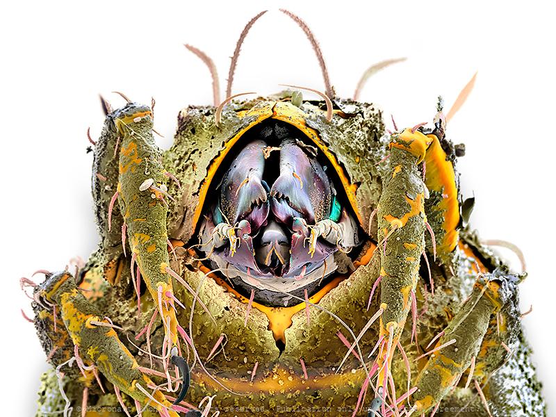
(The Space helmet)
Oribatida: Achipteriidae: Achipteria sp. (a free-living, probably fungivorous or algivorous, soil mite) Achipteridae gehören zu den Hornmilben und ernähren sich je nach Angebot von Pilzen und Algen oder von zerfallendem Laub und Pflanzenmaterial. Oft kommen im Boden Tausende von Individuen pro Quadratmeter vor. Ihre Ausscheidungen werden von Bakterien weiter zersetzt. Die Milben bilden daher einen wichtigen Faktor der Bodenbildung. Die Achipteridae sind wegen ihrer weltweiten Verbreitung und hohen Individuenzahl wichtige Zwischenwirte von Bandwürmern.
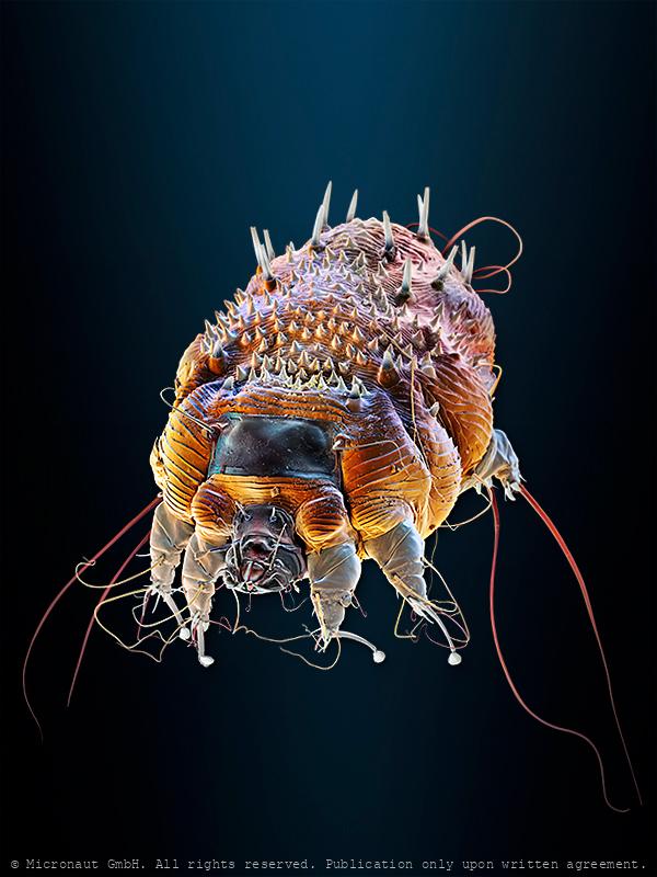
Rabbit Ear mite (Psoroptes cuniculi)
Rabbit Ear Mite (Psoroptes cuniculi): Adults mite grow up to around 0.75mm in length. The legs are long and jointed, bearing suckers on the ends on pairs 1, 2, and 4 in the female and 1, 2, and 3 in the male. Life cycle: egg, larva, two nymph stages, adult. Adults have very characteristic pointed mouthparts. The mucus and fecal material of the parasitic mite induce an inflammatory reaction that leads the rabbit to scratch its ear. The blood that comes out of the scratched lesions serves as a source of nutrition for the parasite. Ear mites are not serious, but if left untreated, infestation can lead to a secondary bacterial infection which can extend to the middle and inner ear causing head tilt, loss of balance, wobbliness (head tilt) and even fatal meningitis. You can tell it is NOT a dust mite because the third pair of legs terminates in long, tassel-like setae. In dust mites, these legs would lack these setae and end in the usual small pad-like 'toes'.
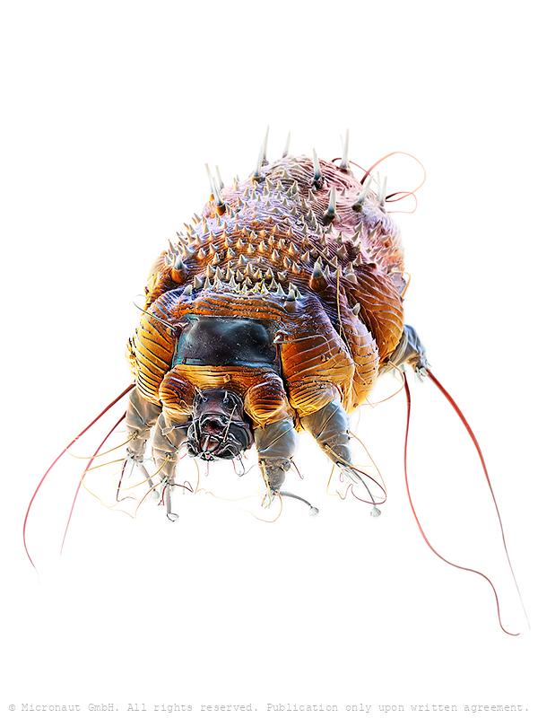
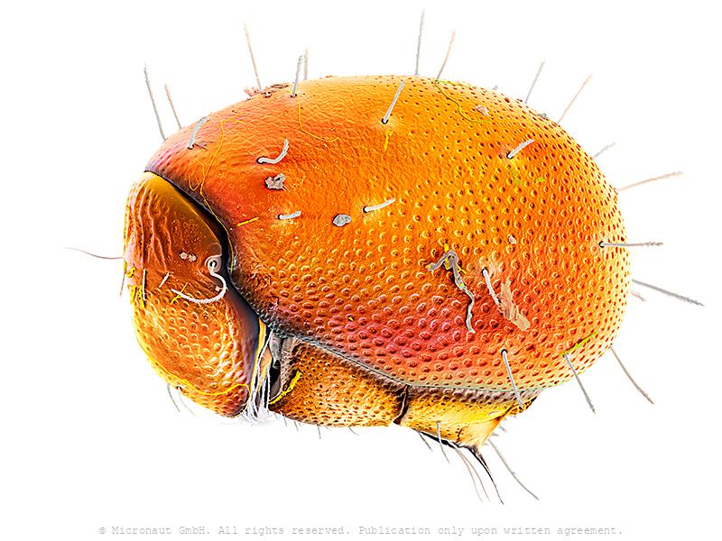
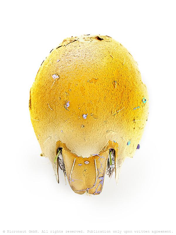
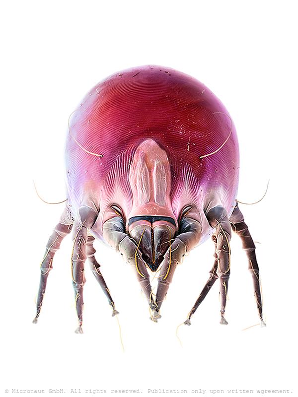
The little devil - House Dust Mite Nr.2 - lateral view (D. farinae)
Hand-colored scanning electron micrograph of a House Dust Mite (D. farinae) by Martin Oeggerli - The house dust mite is a cosmopolitan pyroglyphid mite that lives in human habitation. Dust mites typically measure between 0.2–0.3 millimetres (0.008–0.012 in) in length and feed on organic detritus, such as flakes of shed human skin, and flourish in the stable environment of dwellings. House dust mites are a common cause of asthma and allergic symptoms worldwide. The mite's gut contains potent digestive enzymes (notably proteases) that persist in their feces and are major inducers of allergic reactions such as wheezing. The mite's exoskeleton can also contribute to allergic reactions. There are two different species which occur widely: the European house dust mite (Dermatophagoides pteronyssinus) and the American house dust mite (Dermatophagoides farinae); a third species (Euroglyphus maynei) also occurs worldwide. Unlike scabies mites or skin follicle mites, house dust mites do not burrow under the skin and are not parasitic. Dieses Bild wurde mittels Raster-Elektronen-Mikroskopie aufgenommen und nachträglich manuell koloriert. Es zeigt eine aufgeblasene drohende Hausstaubmilbe.
Predatory Mite (Gamasellus sp.), Nr.1
Colored Scanning Electron Micrograph of a predatory mite (Gamasellus sp.). You can be grateful this animal measures only app. 0.18mm in diameter, because it has a horrible bite: adult Gamasellus eat Springtails (Collembola sp.) of the same size! Compared to juveniles, adults focus on larger prey and eat more often (juvenile: 1 Springtail every 12 days, adult: 1 Springtail every 3 days). Gamasellus is an agile predator and attacks quickly through looping his elongated forelegs over the prey. After this, the chelicerae are moved forward to "kiss" the prey, and inject digestion fluid through a pinhole at the tip of the upper left and right chelicerae. The poisonous fluid tranquilizes and eventually kills and digests the prey. Such a procedure is called pre-oral digestion and it is common in the class of Arachnida, especially in spiders. A multitude of mechanosensory setae ("hairs") are located on the forelegs and pedipalps. They serve the blind predator to monitor the exact position of its prey and make sure it is held in position until the deadly cocktail is effectively working.
House Dust Mite (D. farinae), Nr.1
Hand-colored scanning electron micrograph of a House Dust Mite (D. farinae) by Martin Oeggerli - The house dust mite is a cosmopolitan pyroglyphid mite that lives in human habitation. Dust mites typically measure between 0.2–0.3 millimetres (0.008–0.012 in) in length and feed on organic detritus, such as flakes of shed human skin, and flourish in the stable environment of dwellings. House dust mites are a common cause of asthma and allergic symptoms worldwide. The mite's gut contains potent digestive enzymes (notably proteases) that persist in their feces and are major inducers of allergic reactions such as wheezing. The mite's exoskeleton can also contribute to allergic reactions. There are two different species which occur widely: the European house dust mite (Dermatophagoides pteronyssinus) and the American house dust mite (Dermatophagoides farinae); a third species (Euroglyphus maynei) also occurs worldwide. Unlike scabies mites or skin follicle mites, house dust mites do not burrow under the skin and are not parasitic. Dieses Bild wurde mittels Raster-Elektronen-Mikroskopie aufgenommen und nachträglich manuell koloriert. Es zeigt eine aufgeblasene drohende Hausstaubmilbe.
Pupae of malaria carrying mosquito (Anopheles gambiae)
Mosquito pupae (Anopheles gambiae): Head and thorax are merged into a cephalothorax, with the abdomen curving around underneath. The pupa is comma-shaped and can swim actively by flipping its abdomen, and it is commonly called a "tumbler" because of its swimming action. Mosquitos go through four stages of their life cycle: as with the larva, the pupa of most species must come to the surface frequently to breathe, which they do through a pair of respiratory trumpets on their cephalothoraces. In the course of the metamorphosis, the larva’s eye is subject to drastical differentiation and structural change. While the larva is spending most of its time feeding on algae, bacteria, and other microbes in the surface microlayer and only possess a pit-eye ocellus, the compound eyes of the adult are distinctly separated from one another and much more developed. Similar to other flying insect species, they are the dominant sensory organs for flight orientation.
Soil mite Nr.3 (Eobrachychthonius)
This mite is a Brachychthoniidae and possibly Eobrachychthonius, one of the few oribatids that retain a lateral ocellus that is a simple eye between the seta and bothridium (shown in orange). Eobrachychthonius belongs to the order of the Oribatida, better known as beetle mites or moss mites. Most of them are heavily armored by thick body plates which help to reduce the chance that something eats them. Internally, many are filled with toxins to the same end. Poison dart frogs get their toxins from oribatid mites (and ants), so the defenses don’t always work to their advantage, but often enough for them to have persisted form over 450 million years! Mites secrete an outermost layer of waxes and proteins called the cerotegument (as shown in yellow/green on legs and on remaining body parts). The cerotegument of species found in dry microhabitats has pustulate ornamentation which look a bit like Bucky Balls at high magnification – very similar to some of the types of brochosomes found on Cicadellidae. Likely the cerotegument are water-repellent structures analogous to those found on the upper side of the leaves of water lilies. The 'ear' or 'antenna' is a special seta called a sensillus that can be moved and is sensitive to airborne vibrations. The very deep base (the 'screw-threaded-tube') for the sensillus is called a bothridium. It provides extra support for the sensillus to allow it to vibrate but not to snap off. There is another sensillus and bothridium on the other side of the mite's 'head'. Almost all oribatid species have a pair of sensilli. Unusual for oribatids, Brachychthoniidae can be rather colourful: in the species Eobrachychthonius latior the sclerites are orange in adults and immatures are violet. Suprapleural plates that may be present appear to be infolded, therefore it is not sure if this mite belongs to the genus Eobrachychthonius. The mouthparts on either side of the chelicerae are called pedipalps – homologous to the pincers of sco
Soft tick
Ticks are small arachnids in the order Ixodida, along with mites, constitute the subclass Acarina. Ticks are ectoparasites (external parasites), living by hematophagy on the blood of mammals, birds, and sometimes reptiles and amphibians. Ticks are vectors of a number of diseases, including Lyme disease, Q fever (rare; more commonly transmitted by infected excreta),[1] Colorado tick fever, Rocky Mountain Spotted Fever, tularemia, tick-borne relapsing fever, babesiosis, ehrlichiosis, Tick paralysis and tick-borne meningoencephalitis, as well as bovine anaplasmosis.
Predatory Mite (Gamasellus sp.), Nr.2
Colored Scanning Electron Micrograph of a predatory mite (Gamasellus sp.). You can be grateful this animal measures only app. 0.18mm in diameter, because it has a horrible bite: adult Gamasellus eat Springtails (Collembola sp.) of the same size! Compared to juveniles, adults focus on larger prey and eat more often (juvenile: 1 Springtail every 12 days, adult: 1 Springtail every 3 days). Gamasellus is an agile predator and attacks quickly through looping his elongated forelegs over the prey. After this, the chelicerae are moved forward to "kiss" the prey, and inject digestion fluid through a pinhole at the tip of the upper left and right chelicerae. The poisonous fluid tranquilizes and eventually kills and digests the prey. Such a procedure is called pre-oral digestion and it is common in the class of Arachnida, especially in spiders. A multitude of mechanosensory setae ("hairs") are located on the forelegs and pedipalps. They serve the blind predator to monitor the exact position of its prey and make sure it is held in position until the deadly cocktail is effectively working.
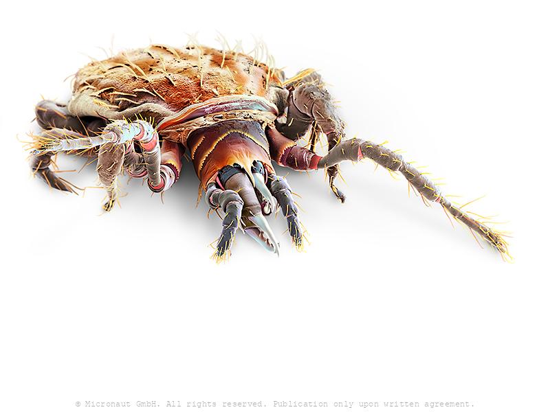
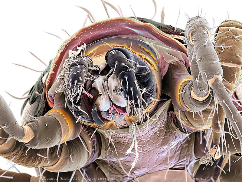
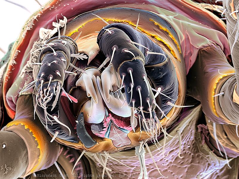
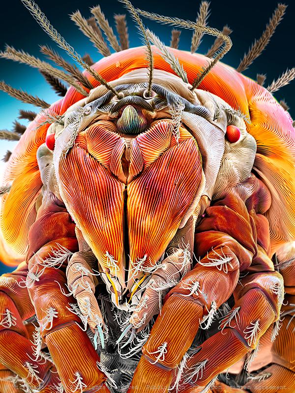
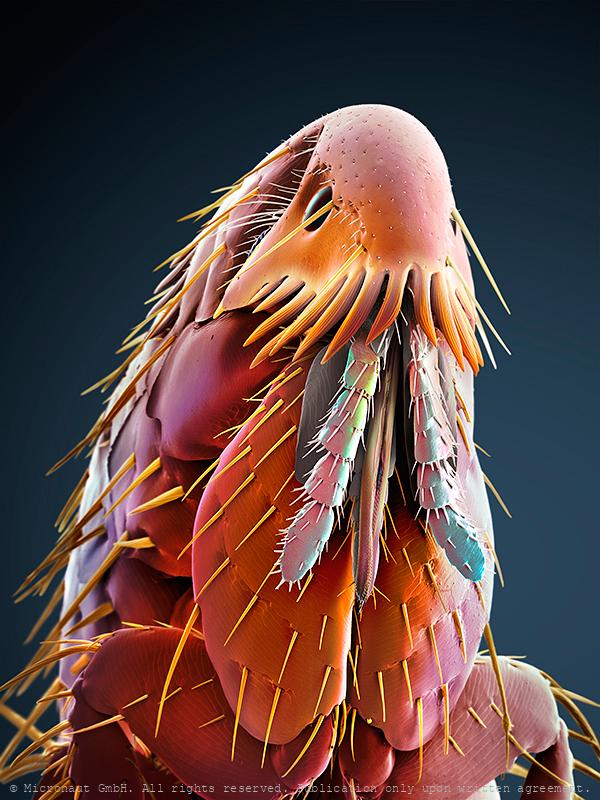
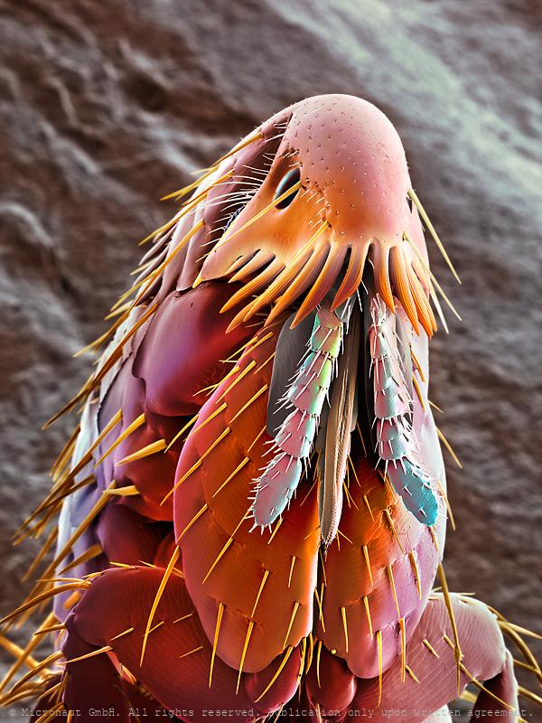
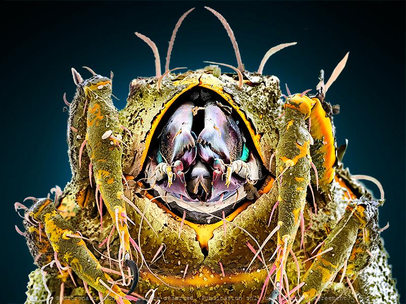
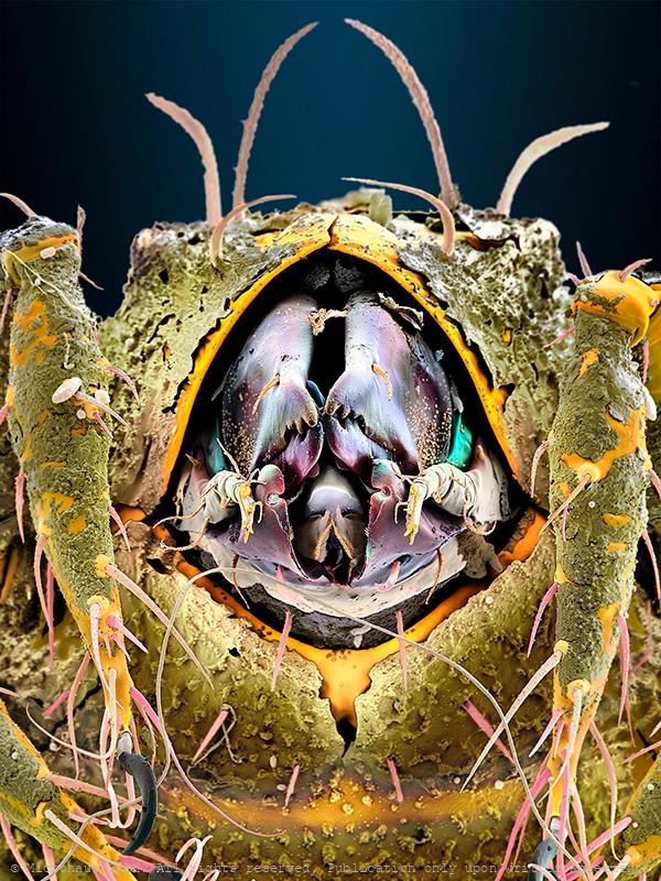
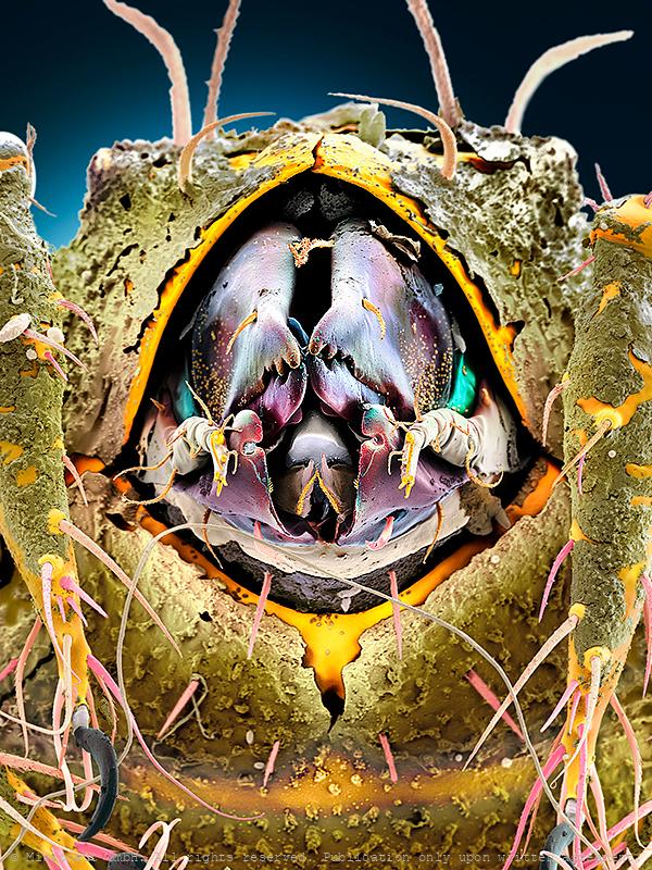
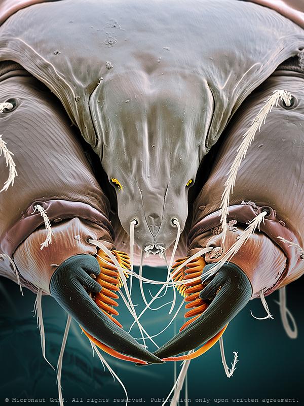
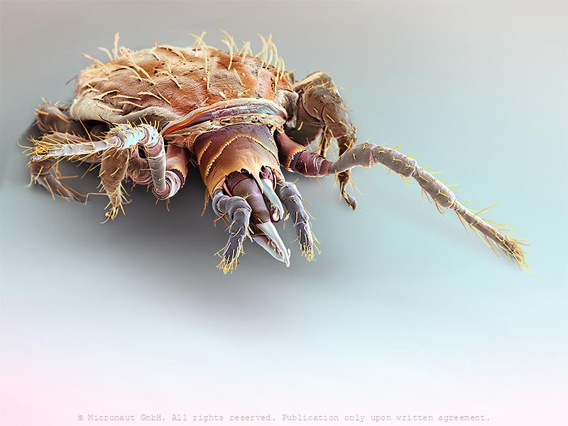
Military Mite - Heavy Duty & the Swiss Pocket Knife
You are looking at the hand-colored scanning-electron-micrograph (SEM) of an oribatid soil mite. The highly magnified portrait exposes a heavily armed creature with a weakly developed head and lots of dirt and humus particles its surface. Most likely this is a member of the Hermanniellidae (Hermanniella sp.). Mite pincers are called chelicera. In this particular case, they are equipped with 3-4 blunt teeth and look very strong. Certainly, this tool has been formed for endurance. Ultra-short blades make it look like a double bolt-cutter: heavy duty! Left and right to the chelicera is a subtile pair of claw-like palps. Below the chelicera and jammed between the palps, you can see a slightly bent pair of scissors. Each blade features a well-shaped notch, right at the tip. In between the scissors the beast holds a golden set of comb-like structures (mostly hidden by the scissors), two superfine pink injection needles and a mysterious pair of bristles. The latter resemble fluffy pipe cleaners. Altogether, the mouth region is exhibiting such a variety of specific tools that it could be the model for a Swiss pocket knife! Of course it would be extremely fascinating to know more about the functional reason of such an incredible set of cutlery, but as it turns out nothing is known about the diet of the beast. Mysterious mites, over and over again! Oribatids often live in extreme environments and can be found from the alpine zone to the marine littoral, on sun-exposed rocks and roofs, sparsely covered by lichens and mosses, as well as in saline soils and salt marshes and in inundation meadows. Furthermore, the Hermanniellidae have an interesting autapomorphy - the tritonymphal cuticle is retained on the notogaster of the adult, so what looks like setae and cuticle on the adult notogaster is actually the scalp of the last nymphal stage. Finally, the individual shows a huge variety of setae, i.e. "bristles", which is a characteristic feature of the family.
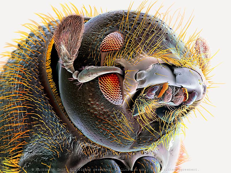
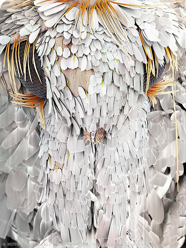
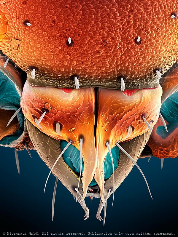
Timber Beetle (Trypodendron lineatum)
Portrait of a Bark Beetle (Spruce & Timber Beetle; Trypodendron lineatum). Bark beetles are a subfamily of the Snout beetles ('Weevils'). 154 species exist within Europe and between 4'000-5'000 worldwide. As primary destruents, they play an essential role in forrest ecosystems, living on dead wood or on dying trees. However, other species are known as notorious pests and are attacking (also) healthy trees. Trypodendron lineatum has a divided compound eye and is a monogamous species and shows brood care. The parents for example clean
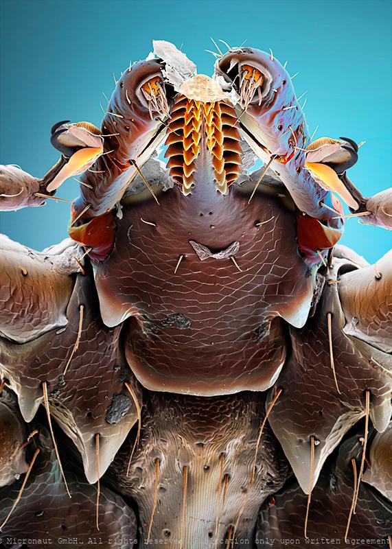
Moth head
Understanding other animals’ sensory abilities allows us to put our capabilities and limitations into perspective and lets us appreciate what we have. It also lets us imagine what it would be like to perceive the world through another creature’s eyes and - inspires technological innovations (ie: when designing robots or night vision goggles). In general the insect compound eye is made up of many separate lenses, called ommatidia. Moths have huge eyes covering almost their entire head. The huge compound eye even lets them see 'in the back of their head' at the cost of a relatively low resolution, because it depends from the number of photoreceptors (i.e. number of lenses) that can be grouped on the relatively small head. Thereby insects perceive the environment as 'patchwork image', as if we would look through the grid of a cellar window. Nevertheless the small moth has a few tricks up their sleeves for a better vision in the dark: (1) Neural summation: moths' brains store up images over time, giving their photoreceptors more chances to see in dim light (our photoreceptors 'stream' images in a constant flow which means old images are almost immediately lost). (2) Ultraviolet photoreceptor type. Instead of red, moths have ultraviolet photoreceoptor types helping them to find flowers at night: in contrast to humans, moths rely on color vision even at very low light without having to switch to achromatic vision to differentiate colors at low intensities. It may give them a less accurate sense of color at daylight, but moths see color at much lower intensities than humans. (3) Antireflective coatings: the ommatidia of moths' are covered with micro-structures which increases light efficiency at night and helps to avoid predators at daylight (frogs, lizards, birds). Anti-reflective coatings have become a key research area in solar panel construction, because they increase the efficiency.
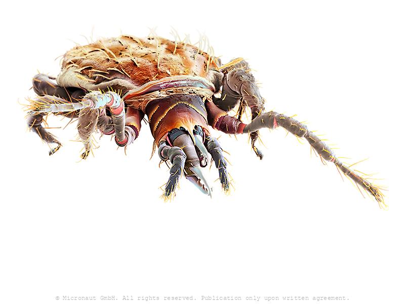
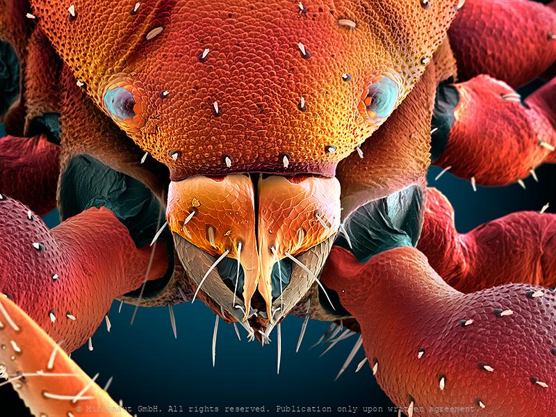
Samurai Armor Protection - The Common Tick (Ixodes ricinus)
Common tick (Ixodes ricinus), hand-colored scanning-electron-micrograph image by science artist Martin Oeggerli. Karuta was a type of armour worn by samurai warriors in Japan. The word karuta comes from the Portuguese word meaning card, (carta) as the small square or rectangular plates that compose the armour resemble traditional Japanese playing cards. In a very similar fashion, the common tick is protected by rigid chitin plates against mechanical damage. Everyone who has ever tried to remove and crush a tick knows very well how tough it is to kill the beast. In addition to the armor-jacket, ticks have also developed other special equipment, e.g. to locate and prepare the right place for the sting (palpi and chelicerae), or to hold on persistently (hypostome and claws), and even resist when the host is moving fast, or is actively trying to remove the parasite by scratching or rubbing. Even more interesting it becomes, if you look at the exquisite details: the barbed stinger (hypostome) features a barrett file structure that is shaped with incomprehensible precision. Each leg ends with a pair of needle-shaped claws, and a fascinating combination of movable splints, adhesion (?) pads, and a mysterious sole which appears to be highly flexible like a towel that can be wrapped around the other structures of the foot. Another high-tech equipment, the Haller's Organ (not visible in this portrait), is located at the front legs and allows the tick to detect chemicals like carbon dioxide, ammonia, and pheromones. It can even sense humidity and infrared light emitted by the warm, blood-filled body a tick wants to find. In-line with the usual allegation of the tick that it can cause infections and spreads diseases (tick-borne encephalitis and/or Lyme disease), this specimen is carrying around a couple of fungi spores and tiny hyphens are sticking to the steel jacket, too. The risk of being bitten by a tick is highest between March and October. Ticks become active a
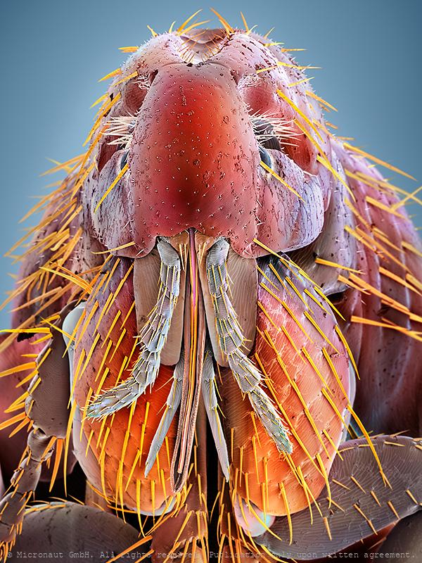
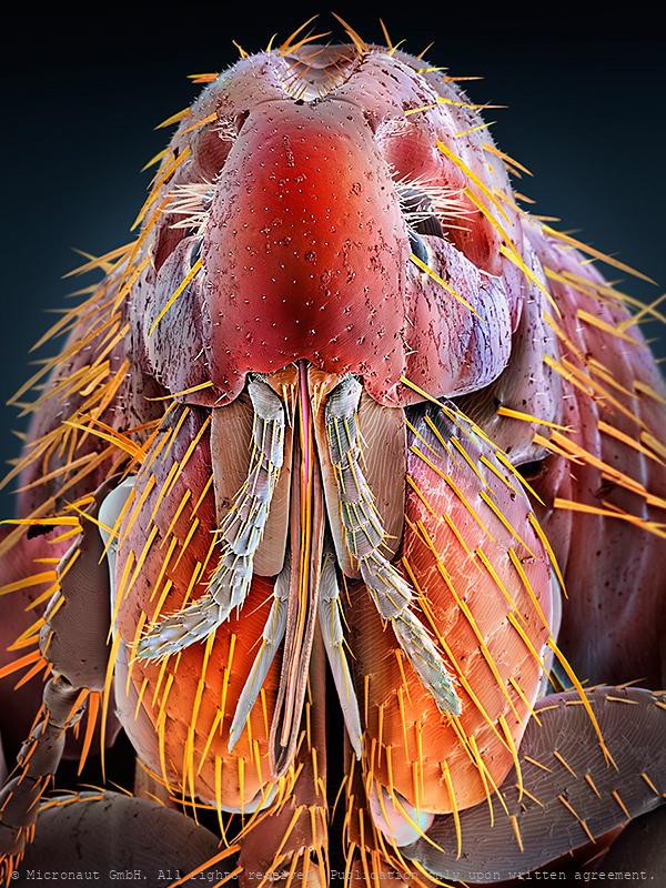
The Lodger - Face Mite (Demodex)
Demodex is a type of mite that lives in human hair follicles, usually on your face. Almost everyone has these mites, but they usually don’t cause problems, except if Demodex can multiply too quickly which is the case in people who are immunocompromised or have other skin conditions. In such a case, face mites can cause an itchy, irritating condition called demodicosis. The parasite is well adapted to its environment through thousands of years of co-evolution with Homo sapiens and extremely tiny (0.15 - 0.4 millimeters). It would take several of them to cover a pin head. Under a microscope, the mite looks slightly transparent and is often covered with scales. It has an elongated body with two segments. The first segment has eight legs and a mouth. When you sleep, the mites come out of your skin’s pores, mate, then go back into your skin to lay eggs. Demodex mites can spread from human to human. Babies aren’t born with mites, but they get mites from the people they live with. Two main species of Demodex live on humans: - Demodex folliculorum: usually lives in smaller hair follicles, especially your eyelashes. They eat skin cells. - Demodex brevis: usually lives near the oil glands in hair follicles. They eat sebum, a greasy substance made by oil glands. This is a photorealistically colored scanning electron micrograph by Martin Oeggerli. The artist also uses colors to make visible specific structures.
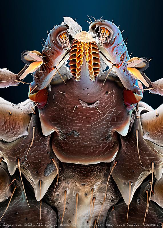
Pseudoscorpion (Chelifer cancroides), Nr.1
Pseudoscorpions are small arachnids with a flat, segmented, pear-shaped body. Despite being harmless and inoffensive creatures, this individual was able to move very quickly to escape. Pseudoscorpions got their name from a pair of huge pincers (pedipalps), resembling those of scorpions. But unlike true Scorpions they have a rounded abdomen that does not extend into a segmented tail with a stinger. The small creatures usually range from 2 to 8 millimetres (0.08 to 0.31 in) in length, with yellowish to red or dark brown colors. Pseudoscorpions are generally beneficial to humans and prey on clothes moth larvae, carpet beetle larvae, booklice, ants, mites, and small flies. Chelifer cancroides is the most commonly found species in homes, where they are often observed in rooms with dusty books. A venom gland and duct are located in the mobile finger of the pincers. The venom is used to capture and immobilize prey. During digestion, a mildly corrosive fluid is poured over the prey to liquify the remains externally before they are ingested. They enter homes by "riding along" attached to insects (known as phoresy). The jaws (chelicera; have a look at the beautiful structure in the center) are used to spin silk which is used to make disk-shaped cocoons for mating, molting, or waiting out cold weather. A comb-like structure (serrula externa), which can be seen on the left side of the chelicera in a close-up picture, is used for grooming. In the center of the chelicera is a membrane called serrula interna. It helps to prevent food and fluids from escaping. Also, two pairs of sensory setae can be found on the chelicera and additional structures which resemble plant pores are located on the cephalothorax (above the eyes in the overview picture). Scientists believe that the latter detect bending and stress on the cuticule and call them lyrifissures. Pseudoscorpions breathe through spiracles on their abdomen (not shown), a trait they share with the insects. Most species have o
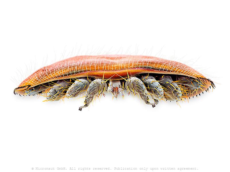
Mite (Speleorchestes sp.)
Mite of the genus Speleorchestes. The genus belongs to the enigmatic Endeostigmata – a perhaps paraphyletic basket that includes the most primitive-looking acariform mites. Endeostigmatans are found throughout the World, but are especially prominent in very dry habitats or dry microhabitats within more mesic ones. Both hot and cold deserts have more than their share of endeostigmatans. This group of mites includes some extraordinarily elongated worm-like species that can be found in the sands of Brazil beaches. However, this particular species is rather small and more mite-like, reaching about a quarter of a millimetre in lenght. Members of the Nanorchestidae also feed on fluids and so their foods are mysterious, although there is one report of a species munching on algae.
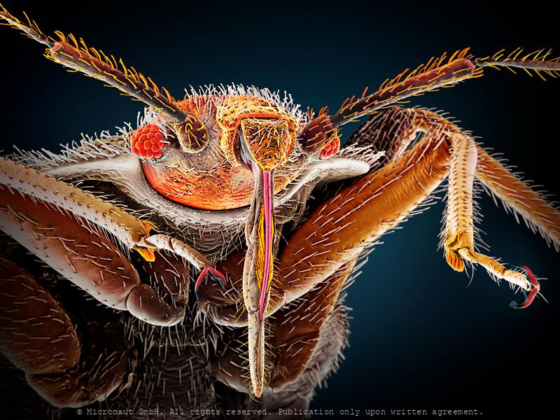
Rat flea (Xenopsylla cheopis)
In general, fleas belong to the insect order Siphonaptera. Their color varies between pale yellow and black and they are rather smalle, usually between 1-8mm long. The heavily sclerotized (armored) body is shiny. The rat flea's primary host is the rat (Rattus norvegicus), but the Oriental rat flea is best known as the vector for the bacteria Yersinia pestis from rats to humans, and for spreading the resulting plague (black death) across Asia and Europe in the middle ages. In the 14th century (between 1347 and 1352), the disease killed 25 million in Europe and between 75 to 100 million worldwide, within five years. Today, the plague is still not wiped out completely and the WHO registers annually between 1'000-3'000 cases worldwide. Like all fleas, Xenopsylla cheopis has mouthparts adapted to cutting through skin and sucking up blood that has pooled. In feeding, it secretes saliva into the wound to prevent the blood from coagulating. Along with the saliva, the flea secretes any bacteria it may have picked up by eating the blood of an infected individual into the host. When Yersinia pestis pathogens enter the gut of the flea, they multiply quickly, blocking food from entering the digestive system. This triggers the hungry flea to bite a new host, further spreading the bacteria. Today the Oriental rat flea can be found in temperate climates, but more often inhabits warm, tropical and subtropical regions, since it needs warm temperatures to pupate. It uses many different mammals as hosts, including rats and humans, and is known also to carry the murine typhus pathogen (Rickettsia typhi) and the mouse and rat tapeworms (Hymenolepis diminut and Hymenolepis nana), the latter can be transmitted when fleas are swallowed by pets or humans.
Catflea (Ctenocephalides felis), Nr.2
Frontansicht eines Katzenflohs. Parasiten sind normalerweise auf einen bestimmten Wirt spezialisiert. Katzenflöhe auf Katzen, nur ausnahmsweise werden auch Menschen gestochen (wenn keine Katze in der nähe ist). Portrait of a cat flea (magn. 158:1 if printed 12.5cm high). The cat flea's primary host is the domestic cat, but this is also the primary flea infesting dogs in most of the world. Cat fleas can transmit other parasites and infections to dogs and cats and also to humans. The most prominent of these are Bartonella, murine typhus, and apedermatitis. The tapeworm Dipylidium caninum can be transmitted when a flea is swallowed by pets or humans. In addition, cat fleas have been found to carry Borrelia burgdorferi, the etiologic agent of Lyme disease, but their ability to transmit the disease is unclear.
Bat flea (ev. Ischnopsyllus sp.)
Coloured scanning electron micrograph (SEM) of a Bat flea, showing its head, mandibles and powerful front legs. This parasite was found on a Daubenton's bat (Myotis daubentonii). The mandibles are used to pierce the host's skin and suck blood. As a result of living on nocturnal hosts Bat fleas have no eyes. Interestingly, Bat fleas have relatively short proboscis, measuring less than 200 µm. This is 33% less compared to the Cat flea (Ctenocephalides felis), 50% less compared to the common Rat flea (Xenopsylla cheopis), and even 90% less compared to the common European Mosquito (Culex pipiens). It remains to be determined if insects that parasitize bats evolved a reduced lenght of their blood sucking instruments due to the thin bat wing membranes that are well supplied with blood vessels and lack subcutaneous fat storage or dense fur. For the author the tiny animal reveals the grace of a flamenco dancer and provides a text-book example for the fascination of invisibly small creatures - and especially for those with a notoriously bad reputation generated in childhood from hearsay and fairy tales. Für den Autor hatte der Floh bereits auf der schwarz-weiss Aufnahme die Grazie einer Flamenco-Tänzerin ausgestrahlt. Nun erkennt man dank den Farben noch einiges mehr und ist gleichzeitig erstaunt wie perfekt die einzelnen Strukturen ineinander passen. Augen hat er keine, er lebt meist in völliger Dunkelheit, dafür sind die Fühler und tastenden Mundwerkzeuge prominent entwickelt und das ganze Gesicht ist äusserst fein behaart. Wenn man nichts sieht und auch kein Echolot hat, dann hilft eben der Tastsinn weiter - wir machen es genauso, falls das Licht einmal nicht funktioniert!
Good night, sleep tight, don't let the bed bugs (Cimex lectulari
Bed bug (Cimex lectularius), hand-colored scanning-electron-micrograph image by science artist Martin Oeggerli. Bed bugs are insects from the genus Cimex that feed on blood, usually at night. Their bites can result in a number of health impacts including skin rashes, psychological effects, and allergic symptoms. Bed bug bite symptoms may take between minutes to days to appear and itchiness is generally present. Some individuals may feel tired or have a fever. Typically, uncovered areas of the body are affected. Sensitivity of humans varies from extreme allergic reaction to no reaction at all (about 20%). However, bed bug bites are not known to transmit any infectious disease. Bed bugs have five immature nymph life stages and a final sexually mature adult stage. They are obligatory bloodsuckers and need at least one blood meal in order to advance to the next stage of development. Special mouth parts allow the parasite to saw through the skin and inject saliva with anticoagulants and painkillers. Bed bug secretions can inhibit the growth of some bacteria and fungi, and antibacterial components from the bed bug could be used against human pathogens, and are considered a possible source of pharmacologically active molecules as a resource for the discovery of new drugs.
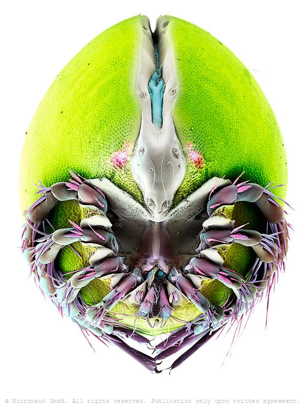
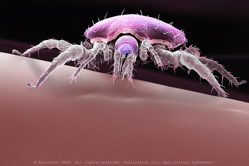
Spidermite eye
Most if not all Acari react to optical stimuli and thus demonstrate photosensitivity, yet not much is known about mite vision. Spider mites have two pairs of relatively large eyes. They can be used for color vision and to detect near-ultraviolet light waves.The pair of eyes on each side of Tetranychus urticae consists of fifteen retinular cells, six pigment cells, six corneal cells, and one vitreous cell. Responses of the summer form of adult females placed in near-ultraviolet and green light have shown that two separate photoreceptor systems are present and are able to discriminate between upper and lower leaf surface. Spider mites rely on vision to locate the preferred habitate, avoid deleterious UV-radiation, and to find a place to hide.
Military Mite - Heavy Duty & the Swiss Pocket Knife
You are looking at the hand-colored scanning-electron-micrograph (SEM) of an oribatid soil mite. The highly magnified portrait exposes a heavily armed creature with a weakly developed head and lots of dirt and humus particles its surface. Most likely this is a member of the Hermanniellidae (Hermanniella sp.). Mite pincers are called chelicera. In this particular case, they are equipped with 3-4 blunt teeth and look very strong. Certainly, this tool has been formed for endurance. Ultra-short blades make it look like a double bolt-cutter: heavy duty! Left and right to the chelicera is a subtile pair of claw-like palps. Below the chelicera and jammed between the palps, you can see a slightly bent pair of scissors. Each blade features a well-shaped notch, right at the tip. In between the scissors the beast holds a golden set of comb-like structures (mostly hidden by the scissors), two superfine pink injection needles and a mysterious pair of bristles. The latter resemble fluffy pipe cleaners. Altogether, the mouth region is exhibiting such a variety of specific tools that it could be the model for a Swiss pocket knife! Of course it would be extremely fascinating to know more about the functional reason of such an incredible set of cutlery, but as it turns out nothing is known about the diet of the beast. Mysterious mites, over and over again! Oribatids often live in extreme environments and can be found from the alpine zone to the marine littoral, on sun-exposed rocks and roofs, sparsely covered by lichens and mosses, as well as in saline soils and salt marshes and in inundation meadows. Furthermore, the Hermanniellidae have an interesting autapomorphy - the tritonymphal cuticle is retained on the notogaster of the adult, so what looks like setae and cuticle on the adult notogaster is actually the scalp of the last nymphal stage. Finally, the individual shows a huge variety of setae, i.e. "bristles", which is a characteristic feature of the family.
Pseudoscorpion (Chelifer cancroides), Nr.2
Pseudoscorpions are small arachnids with a flat, segmented, pear-shaped body. Despite being harmless and inoffensive creatures, this individual was able to move very quickly to escape. Pseudoscorpions got their name from a pair of huge pincers (pedipalps), resembling those of scorpions. But unlike true Scorpions they have a rounded abdomen that does not extend into a segmented tail with a stinger. The small creatures usually range from 2 to 8 millimetres (0.08 to 0.31 in) in length, with yellowish to red or dark brown colors. Pseudoscorpions are generally beneficial to humans and prey on clothes moth larvae, carpet beetle larvae, booklice, ants, mites, and small flies. Chelifer cancroides is the most commonly found species in homes, where they are often observed in rooms with dusty books. A venom gland and duct are located in the mobile finger of the pincers. The venom is used to capture and immobilize prey. During digestion, a mildly corrosive fluid is poured over the prey to liquify the remains externally before they are ingested. They enter homes by "riding along" attached to insects (known as phoresy). The jaws (chelicera; have a look at the beautiful structure in the center) are used to spin silk which is used to make disk-shaped cocoons for mating, molting, or waiting out cold weather. A comb-like structure (serrula externa), which can be seen on the left side of the chelicera in a close-up picture, is used for grooming. In the center of the chelicera is a membrane called serrula interna. It helps to prevent food and fluids from escaping. Also, two pairs of sensory setae can be found on the chelicera and additional structures which resemble plant pores are located on the cephalothorax (above the eyes in the overview picture). Scientists believe that the latter detect bending and stress on the cuticule and call them lyrifissures. Pseudoscorpions breathe through spiracles on their abdomen (not shown), a trait they share with the insects. Most species have o
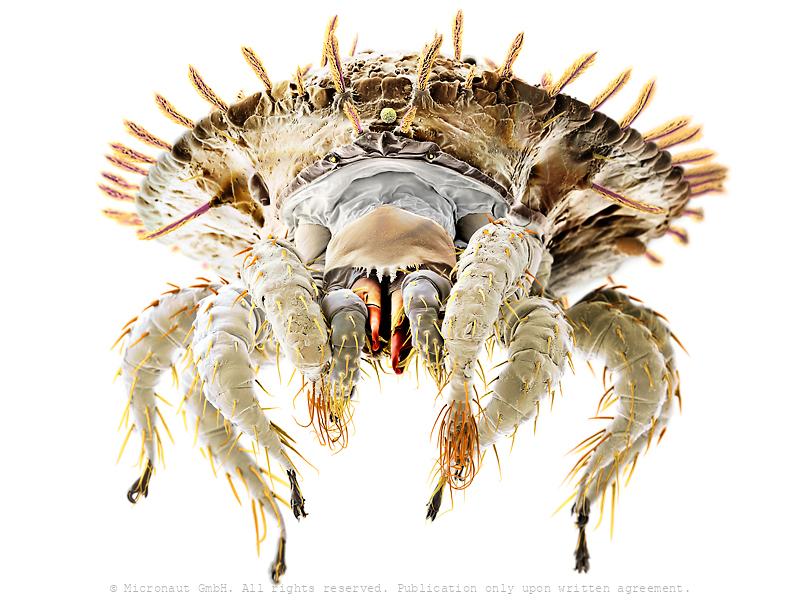
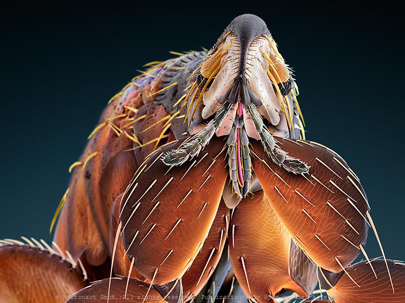
Mite (Cheyletidae, Leach 1815)
Title: The Superparasite (Cheyletid sp.) / Der Superparasit (Cheyletid sp.) Beschreibung: Als Superparasit bezeichnet man eine Art (in diesem Fall eine Milbe), welche sich von Parasiten (in diesem Fall andere Milbenarten) ernährt und diese lästigen Biester beseitigt - also aus der Sicht des Wirtes (in diesem Fall ein Specht) ein höchst willkommener Gast. Der Superparasit verfügt über eine Reihe von hochspezialisierten Mundwerkzeugen - vergleichbar mit einem Schweizer Taschenmesser, um seine Beute zu packen, zu immobilisieren, zu verspeisen, oder um sich zu putzen, zu paaren, etc. Dieses Werk stammt aus einer Serie von Milben-Portraits, welche die ausserordentliche Vielfalt dieser Tiergruppe aufzeigt und an den ‘International Photography Awards’ 2011 mit dem 1. Preis im Bereich Spezial-Fotografie ausgezeichnet wurde. Captions: This looks like the cheyletid from Chrysocolaptes lucidus (a woodpecker) from the Philippines. It is predatory and probably feeds on other mites in the plumage of the bird or in its nest. The eyes are a darker red, but are not shown in this picture. The creature also shows a large white patch in the middle of the body but that’s just the excretory glad filled up with guanine crystals, and is ‘optional’ (as it wouldn’t be there after the mite excretes). The things colored in black are the palpal claws. The structures in orange and yellow are two pairs of highly modified setae. All three pairs of structures are used for sensing and manipulating prey. The bird and its parasite have been found on an expedition in 1972 in the Philippines, and were stored in the deep freezer of a candian museum collection until 2011. Then, this specimen was collected, fixed in alcohol and shipped to Switzerland to be analyzed and illustrated by the author (Martin Oeggerli), utilizing a scanning electron microscope (SEM). This work is part of a the artist’s long-term project, i.e. the series of mite portraits which has become famous through th
Samurai Armor Protection - The Common Tick (Ixodes ricinus)
Common tick (Ixodes ricinus), hand-colored scanning-electron-micrograph image by science artist Martin Oeggerli. Karuta was a type of armour worn by samurai warriors in Japan. The word karuta comes from the Portuguese word meaning card, (carta) as the small square or rectangular plates that compose the armour resemble traditional Japanese playing cards. In a very similar fashion, the common tick is protected by rigid chitin plates against mechanical damage. Everyone who has ever tried to remove and crush a tick knows very well how tough it is to kill the beast. In addition to the armor-jacket, ticks have also developed other special equipment, e.g. to locate and prepare the right place for the sting (palpi and chelicerae), or to hold on persistently (hypostome and claws), and even resist when the host is moving fast, or is actively trying to remove the parasite by scratching or rubbing. Even more interesting it becomes, if you look at the exquisite details: the barbed stinger (hypostome) features a barrett file structure that is shaped with incomprehensible precision. Each leg ends with a pair of needle-shaped claws, and a fascinating combination of movable splints, adhesion (?) pads, and a mysterious sole which appears to be highly flexible like a towel that can be wrapped around the other structures of the foot. Another high-tech equipment, the Haller's Organ (not visible in this portrait), is located at the front legs and allows the tick to detect chemicals like carbon dioxide, ammonia, and pheromones. It can even sense humidity and infrared light emitted by the warm, blood-filled body a tick wants to find. In-line with the usual allegation of the tick that it can cause infections and spreads diseases (tick-borne encephalitis and/or Lyme disease), this specimen is carrying around a couple of fungi spores and tiny hyphens are sticking to the steel jacket, too. The risk of being bitten by a tick is highest between March and October. Ticks become active a
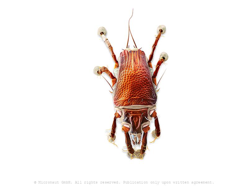
Watermite (Hydrachna sp.), Nr.1
Prostigmata: Hydrachnidae: Hydrachna sp. (a water mite predatory on the eggs of aquatic Heteroptera). With long swimming hairs on rear legs.
Feather mites (Dicamaralges loricatus) from Woodpecker (Chrysocolaptes lucidus)
These mites are Dicamaralges loricatus (Vitzthum) (Acari: Astigmata: Psoroptoididae), feather mites from the woodpecker Chrysocolaptes lucidus (Picidae) from the Philippines. The big mite is the male and the small one is a juvenile female. They are not mating, but rather the male is guarding the young female until she moults into an adult, and then he will copulate with her. In life, the body colour is a light, shiny golden brown (http://simple.wikipedia.org/wiki/Bronze_(color))
The First Dsease of Humans (Scabies, 1687)
This is a hand colored scanning electron micrograph (SEM) of Sarcoptes scabiei, the itch mite, which is a parasitic arthropod burrowing into human skin, thereby causing scabies. The discovery of the itch mite in 1687 marked scabies as the first disease of humans with a known cause. Other mammals can also become infected, including dogs, cats, ungulates, wild boars, bovids, wombats, koalas, and great apes. About 2% of the British population is thought to be infected with these mites, which take about 25 minutes to an hour to burrow into the skin. The burrowing is carried out using the mouth parts and special cutting surfaces on the front legs. A burrowing mite anchors with suckers on its feet, furrows and spines, located on its body surface. Eggs are laid in small numbers as the mite burrows. As these hatch, six-legged larvae climb out on to the skin and search for hair follicles, where they feed and develop. In the hair follicles, the larvae show the first nymphal stages, with eight legs.
Pupae of malaria carrying mosquito (Anopheles gambiae)
Mosquito larvae have well developed mouthparts, which are used to induce a current and feed from algae and bacteria. Mosquitos go through four stages of their life cycle: egg, larvae, pupae and imago (adult insect). Adult females lay their eggs in standing water. The first three stages are aquatic and last 5–14 days, depending on the species and temperature. The pupa is comma-shaped, as in Anopheles when viewed from the side, and is commonly called a "tumbler". As with the larvae, pupae must come to the surface frequently to breathe, which they do through a pair of respiratory trumpets on the cephalothorax. After a few days, the pupa rises to the water surface, the dorsal surface of the cephalothorax splits and the adult mosquito emerges.
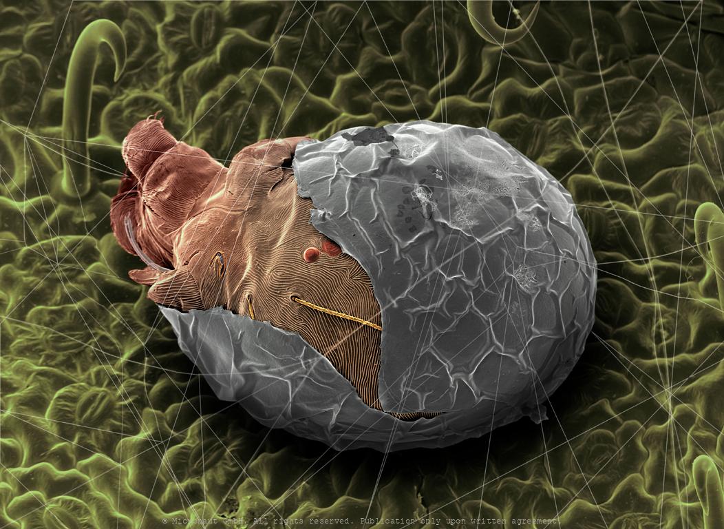
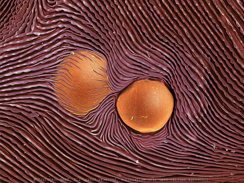
Hatching spider mite
Hatching spider mite
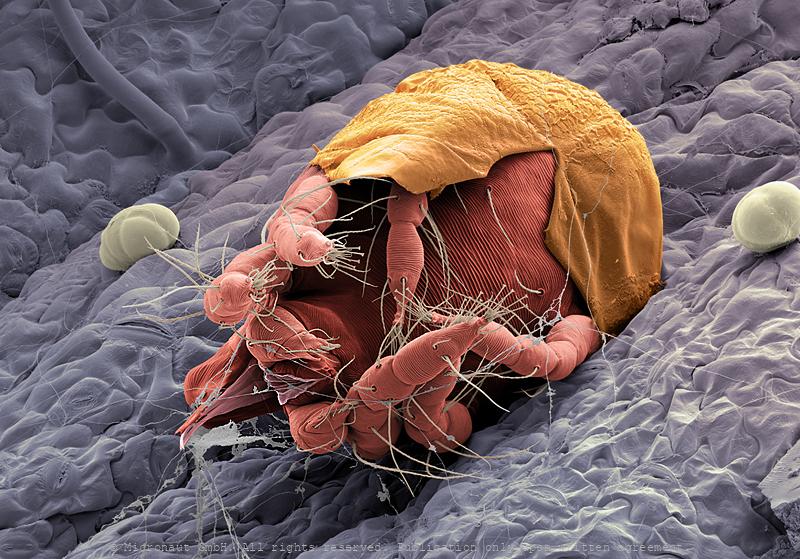
Hatching spider mite
Hatching spider mite
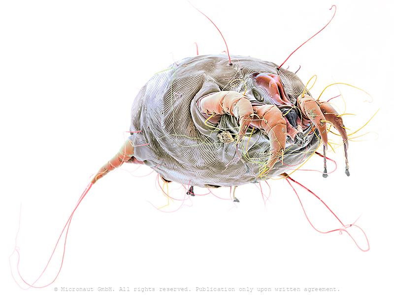
Parasitic mite (unknown species)
Predatory Mite (Gamasellus sp.), Nr.1
Colored Scanning Electron Micrograph of a predatory mite (Gamasellus sp.). You can be grateful this animal measures only app. 0.18mm in diameter, because it has a horrible bite: adult Gamasellus eat Springtails (Collembola sp.) of the same size! Compared to juveniles, adults focus on larger prey and eat more often (juvenile: 1 Springtail every 12 days, adult: 1 Springtail every 3 days). Gamasellus is an agile predator and attacks quickly through looping his elongated forelegs over the prey. After this, the chelicerae are moved forward to "kiss" the prey, and inject digestion fluid through a pinhole at the tip of the upper left and right chelicerae. The poisonous fluid tranquilizes and eventually digests the prey. Such a procedure is called pre-oral digestion and it is common in the class of Arachnida, especially in spiders. A multitude of mechanosensory setae ("hairs") are located on the forelegs and pedipalps. They serve this blind predator as a monitor to determine the exact position of the prey and makes sure it is held in position until the deadly cocktail is effectively working.
(The Space helmet)
Oribatida: Achipteriidae: Achipteria sp. (a free-living, probably fungivorous or algivorous, soil mite) Achipteridae gehören zu den Hornmilben und ernähren sich je nach Angebot von Pilzen und Algen oder von zerfallendem Laub und Pflanzenmaterial. Oft kommen im Boden Tausende von Individuen pro Quadratmeter vor. Ihre Ausscheidungen werden von Bakterien weiter zersetzt. Die Milben bilden daher einen wichtigen Faktor der Bodenbildung. Die Achipteridae sind wegen ihrer weltweiten Verbreitung und hohen Individuenzahl wichtige Zwischenwirte von Bandwürmern.