Microcosm I (animals)
This is the place of the earth’s smallest living organisms and their structures: concealed by sheer size, various forms of life spend their just below the radar of the human eye. Micronaut collects and explores invisibly small creatures and their structures from every corner of the earth. All images are based on Scanning-Electron-Microscopy (SEM). By utilizing electrons instead of photons, SEM technology is capable to produce magnifications up to 500.000-times or even more, thereby expanding the limits of traditional photography.
To reconstruct the colors -which cannot be captured through SEM technology- the pictures are manually colored (post-processing). The manual work combines Dr. Oeggerli’s scientific knowledge and his artistic eye, creating new insights into scientific phenomena and unknown territory. The following two projects contain selected images of animals.
«Martin has put scientific photography on a new esthetic level.»
Wolfgang Bengel, MD
Vice president DGZMB, Lennart Nilsson-Award Committee Member
Micronaut images are rights-managed. If you want to get a quote, please contact us, providing the following information: (1) image name, (2) specific use, (3) industry, (4) distribution area, (5) format, (6) circulation or print run, and (7) duration.
Please note that we cannot answer incomplete requests. Thank you.
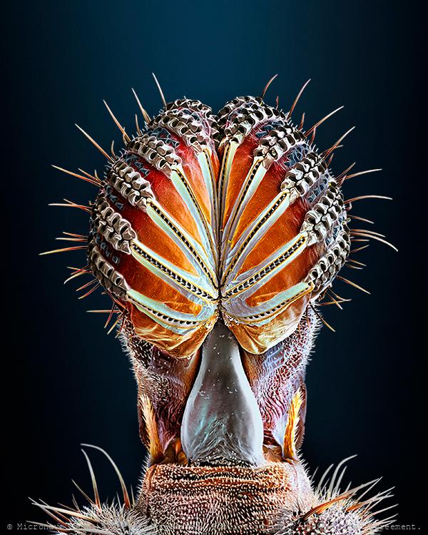
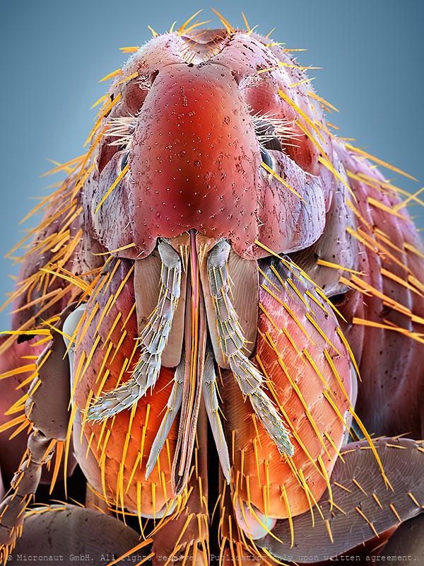
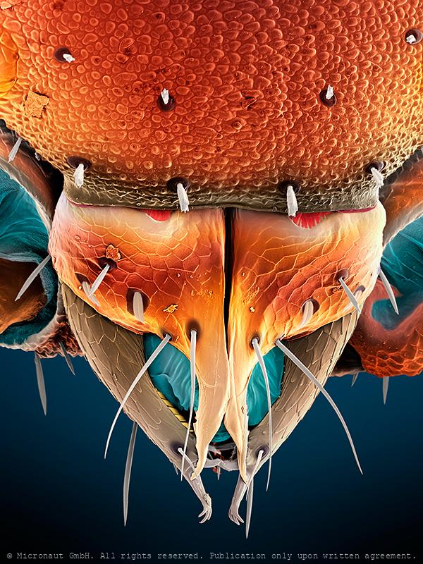
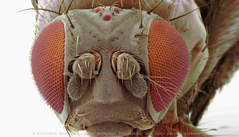
Hyper realistic fly proboscis (Drosophila melanogaster)
Hyper realistic fly (Drosophila melanogaster) proboscis. This highly enlarged microcraph is based on Scanning-Electron-Microscopy, and was created and manually colored by Martin Oeggerli (Micronaut). His realistic paintings shed light on invisibly small matters. By dramatically enlarging and exposing his tiny and unfamiliar subjects, Martin Oeggerli creates a surrealistic tactility of subjects we can neither see, nor touch. Fruitflies possess mouthparts adapted for sucking and feed on plant juices. The tip of the proboscis in some flies has spongy structures which conveys liquid food to the mouth. Solid foods are dissolved in a drop of saliva & sucked up in the same way.
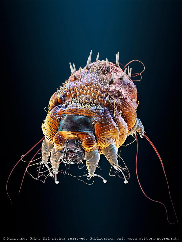
Scanning electron microscope image of mosquito larva (family Culicidae) mouth parts.
Scanning electron microscope image of the mouth parts of a mosquito larva (family Culicidae). The collection of hairs (light brown) are feeding structures used to filter water. The hairs beat through the water filtering out algae, bacteria and other micro-organisms that the larva feeds on.
Pupae of malaria carrying mosquito (Anopheles gambiae)
Mosquito larvae have well developed mouthparts, which are used to induce a current and feed from algae and bacteria. Mosquitos go through four stages of their life cycle: egg, larvae, pupae and imago (adult insect). Adult females lay their eggs in standing water. The first three stages are aquatic and last 5–14 days, depending on the species and temperature. The pupa is comma-shaped, as in Anopheles when viewed from the side, and is commonly called a "tumbler". As with the larvae, pupae must come to the surface frequently to breathe, which they do through a pair of respiratory trumpets on the cephalothorax. After a few days, the pupa rises to the water surface, the dorsal surface of the cephalothorax splits and the adult mosquito emerges.
Mosquito egg surface
Adult females lay their eggs in standing water, which can be a salt-marsh, a lake, a puddle, a natural reservoir on a plant, or an artificial water container such as a plastic bucket. Usually many eggs are clumped together (i.e. 'egg rafts'). Each egg has a hydrophobic surface: a thin layer of air is trapped by 'tent-like' micro-structures, to avoid sinking of the egg/egg-raft.
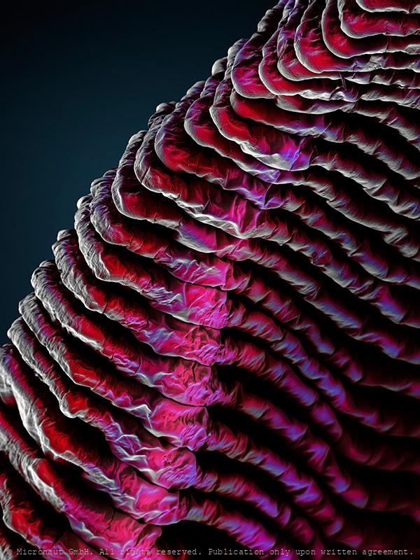
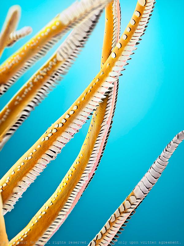
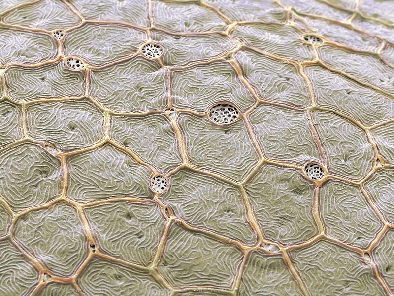
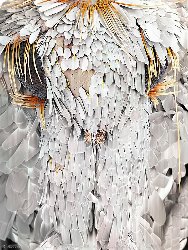
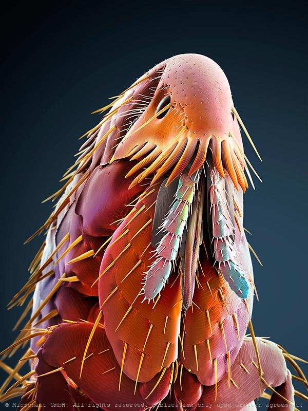
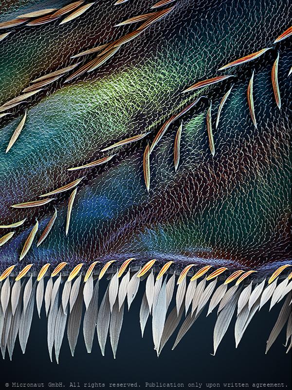
Penguin feather
Penguin feather fine structures, hand-colored Scanning-Electron-Micrograph by Martin Oeggerli / Micronaut.

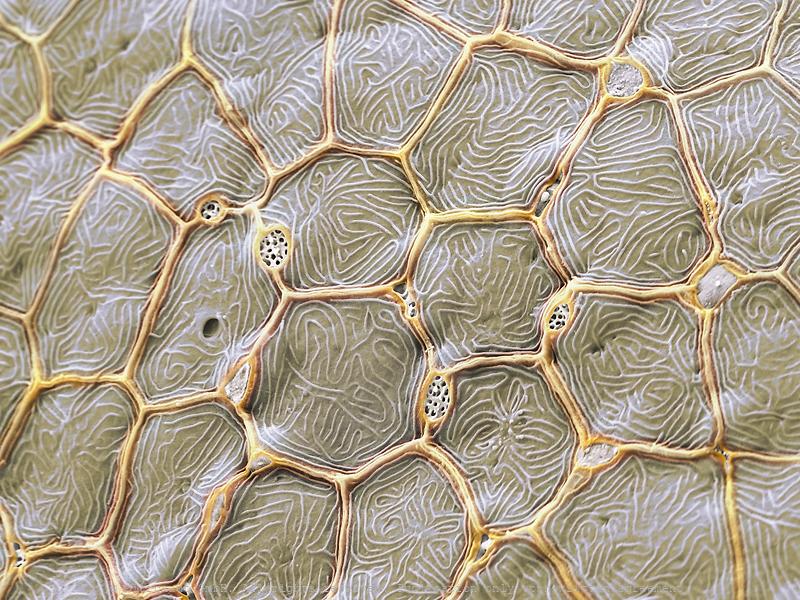
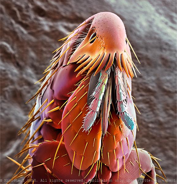
Penguin feather
Penguin feather fine structures, hand-colored Scanning-Electron-Micrograph by Martin Oeggerli / Micronaut.
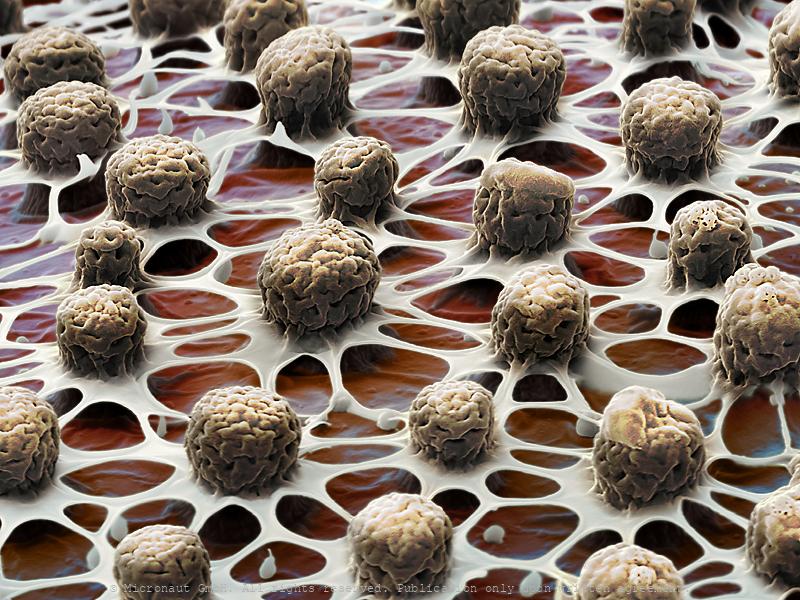
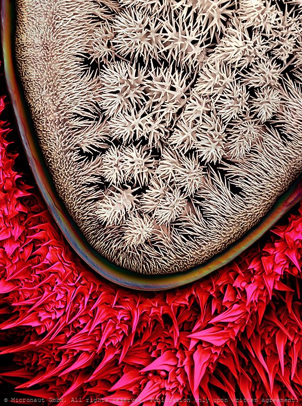
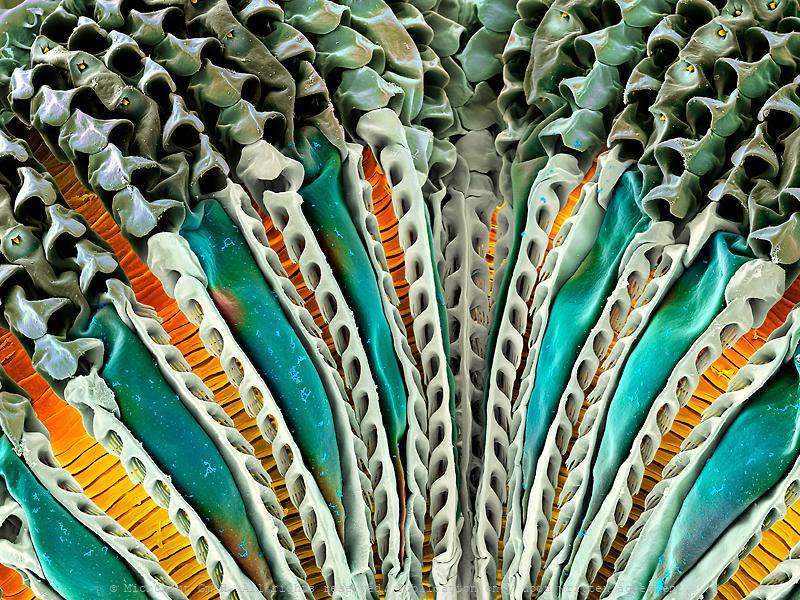
Lung of a Ghost - Gills of a Shrimp
The lungs of a shrimp, called gills, as the only respiratory organ of the animal, and one of the main organs involved in the osmotic adjustment process . Due to their delicate nature and exposure to substances, pathogens and toxins, gills are one of the most important organs for disease detection in shrimps. Artwork created by Martin Oeggerli (Micronaut). The picture is based on scanning-electron-microscopy technology, and all colors were manually added to visualize the gills of a shrimp which are enclosed in the branchial chamber difficult to observe with standard technology.
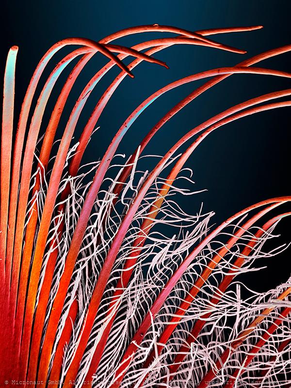
Sensory hairs of a shrimp
Leg hairs (called setae) of a shrimp (crustacea), hand-colored scanning-electron-micrograph by Martin Oeggerli (Micronaut). Most rustaceans (e.g. lobster, crabs, and shrimp) have an extensive array of sensory hairs covering their bodies and they have many different purposes. The setae are the tiny hairs on the legs of the shrimp. Setae can be used to filter food through the water and push it toward the mouth. Some shrimp use the setae to incubate fertilized eggs, and others use setae for grooming. Furthermore, the setae can be used to detect movement in the water, detecting current changes and water flow. It has been reported that some species have also developed highly specialized setae for chemosensory communication. Others use setae to detect water- and substrate-born vibrations and sound as part of their intraspecific communication. For example the sensory hairs (setae) located on the claws of the Australian freshwater crayfish are sensitive to water vibration frequencies between 150-300Hz. This picture shows the tips of setae which are arranged at the very tip of the legs of a freshwater shrimp (unknown species).
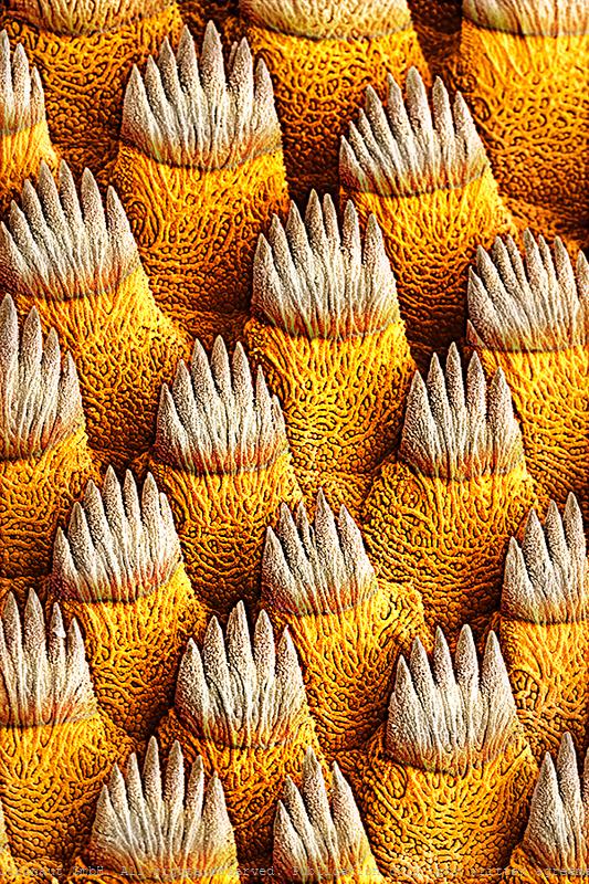
Zebrafish scales (Danio rerio), Nr.1
Zebrafish scales. Coloured scanning electron micrograph (SEM) of zebrafish scales (Danio rerio) scales. Fish scales are are classified by shape. As the fish grows, the scale increase in size and growth rings called circuli become visible. During cooler months of the year the scale grows more slowly and the circuli are closer together leaving a band called an annulus. By counting the annuli it is possible to estimate the age of the fish.
Zebrafish scales (Danio rerio), Nr.2
Zebrafish scales. Coloured scanning electron micrograph (SEM) of zebrafish scales (Danio rerio) scales. Fish scales are are classified by shape. As the fish grows, the scale increase in size and growth rings called circuli become visible. During cooler months of the year the scale grows more slowly and the circuli are closer together leaving a band called an annulus. By counting the annuli it is possible to estimate the age of the fish.
Hirudino medicinalis surface
Hirudino medicinalis surface
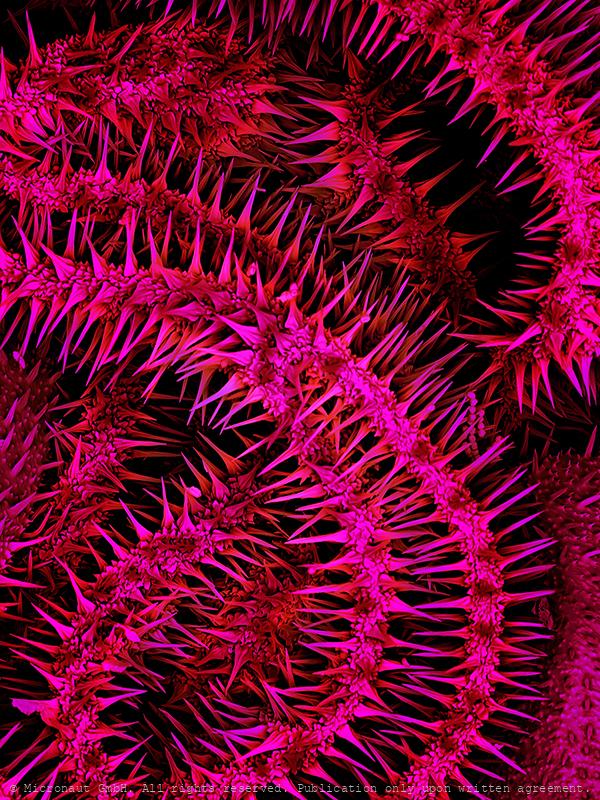
Suction Cup (Caterpillar foot)
Colored scanning electron micrograph (SEM) of a caterpillar foot (called proleg), co-produced with Sheri Neva, 2021. Insects have three pairs of legs which are located close to the head of the caterpillar. In addition, caterpillars are equipped with a number of fleshy prolegs further along the body. The prolegs help caterpillars to walk and get a grip, even on slippery surfaces. The proleg may be equipped with several rows of hooks (not in this species), and/or microscopically small hairs. These hairs enable the gecko to cling to smooth surfaces by taking advantage of weak intermolecular forces, known as Van der Waals forces, similar to the Gecko. The caterpillars of some butterfly species further produce threads of silk to secure and increase their steep climbing routes, and eventually also to produce the cocoon and attach it safely to a twig or leaf during metamorphosis. The silk of the Silkworm (caterpillar) is commercially woven into cloth.
Caterpillar Surface
Coloured scanning electron micrograph (SEM) of caterpillar skin. Insects are often equipped with exotic surface structure like urticating hairs, thorns, or hooks.
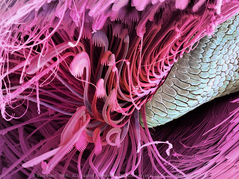
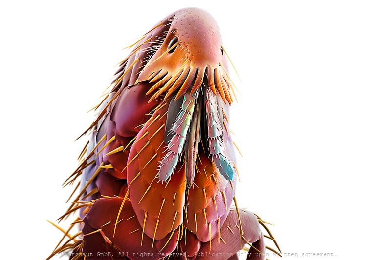
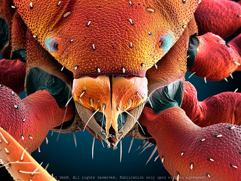
Fly proboscis (Drosophila melanogaster)
Coloured scanning electron micrograph (SEM) of the tip of the proboscis (snout) of a fruitfly (order Diptera). Fruitflies possess mouthparts adapted for sucking and feed on plant juices. The tip of the proboscis in some flies has spongy structures which conveys liquid food to the mouth. Solid foods are dissolved in a drop of saliva & sucked up in the same way.
Collembola Snout
Springtail (Collembola sp.) mouth region. Springtails are found in leaf litter and soil. They have internal mouthparts and are no longer considered to be insects (which have external mouthparts). Collembola lack a tracheal respiration system, which forces them to respire through a porous cuticle (see ornamented surface structure). In sheer numbers, Springtails are among the most abundant organisms. With estimates of 100'000 per cubic metre of topsoil, they are essentially everywhere on Earth where soil and related habitats occur. Springtails can be found essentially everywhere on Earth where soil and related habitats occur. With insatiable appetite they eat their way through the topsoil and thereby produce what is essential for plants: excrements rich of essential nutrients that would otherwise be inaccessible for plants. Their stomach breaks down the cellulose from leaves and stems. Springtails are the favourite meal for predators such as mites and pseudo scorpions. To get away from them, they possess a kind of spring under their abdomen, their “springtail”. It can be moved like a folding knife in a fraction of a second. The force hurls the animal several centimetres away, provided there is enough space.
Velcro claw (Coleopterus sp.)
Claw of a beetle (Coleoptera, Cerambycidae). Coloured scanning electron micrograph (SEM) of hairs and claw on the underside of a beetle's foot (family Cerambycidae). These microscopic hairs enable the beetle to cling to very smooth surfaces such as even windows and ceilings.
Spider Silk, Nr.1
Spider silk is a protein fibre spun by spiders. Spiders use their silk to make webs or other structures, which function as sticky nets to catch other animals, or as nests or cocoons to protect their offspring, or to wrap up prey. They can also use their silk to suspend themselves, to float through the air, or to glide away from predators. Most spiders vary the thickness and stickiness of their silk for different uses. A single spider can produce up to seven different types of silk for different uses. Consisting of mainly protein, silks are about a sixth of the density of steel (1.3 g/cm3). As a result, a strand long enough to circle the Earth would weigh less than 500 grams (18 oz). Making the silk acidic (pH 4) is a protection from fungi and bacteria that would otherwise digest the protein. The spider silk shown here results from combination of four fibres. This hand-painted picture was produced using a scanning electron microscope, by Micronaut (Martin Oeggerli).
Spider Silk, Nr.2
Spider silk is a protein fibre spun by spiders. Spiders use their silk to make webs or other structures, which function as sticky nets to catch other animals, or as nests or cocoons to protect their offspring, or to wrap up prey. They can also use their silk to suspend themselves, to float through the air, or to glide away from predators. Most spiders vary the thickness and stickiness of their silk for different uses. A single spider can produce up to seven different types of silk for different uses. Consisting of mainly protein, silks are about a sixth of the density of steel (1.3 g/cm3). As a result, a strand long enough to circle the Earth would weigh less than 500 grams (18 oz). Making the silk acidic (pH 4) is a protection from fungi and bacteria that would otherwise digest the protein. The spider silk shown here results from combination of four fibres. This hand-painted picture was produced using a scanning electron microscope, by Micronaut (Martin Oeggerli).
Spider Silk, Nr.1
Spider silk is a protein fibre spun by spiders. Spiders use their silk to make webs or other structures, which function as sticky nets to catch other animals, or as nests or cocoons to protect their offspring, or to wrap up prey. They can also use their silk to suspend themselves, to float through the air, or to glide away from predators. Most spiders vary the thickness and stickiness of their silk for different uses. A single spider can produce up to seven different types of silk for different uses. Consisting of mainly protein, silks are about a sixth of the density of steel (1.3 g/cm3). As a result, a strand long enough to circle the Earth would weigh less than 500 grams (18 oz). Making the silk acidic (pH 4) is a protection from fungi and bacteria that would otherwise digest the protein. The spider silk shown here results from combination of four fibres. This hand-painted picture was produced using a scanning electron microscope, by Micronaut (Martin Oeggerli).
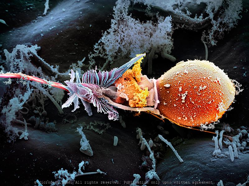
The Magic Bullet - Cnidocyte
The name Cnidaria comes from the Greek word "cnidos", which means stinging nettle. Invisibly small, the cnidocyst which are responsible for the explosive pain caused by jellyfish, remains a mysterious weapon until today. Cnidarians are a varied group of animals including corals, sea anemones, sea pens, sea pansies, jellyfishes, and hydroids. They range among the most beautiful of all marine invertebrates. Many species have a flower-like look and are often considered delicate and soft. But they possess a well-concealed yet potent arsenal of harpoon-like stinging cells. Stings from several species can be very painful, and in some cases, even fatal. Box jellyfish and sea wasps represent the biggest “stinging” threat to divers and swimmers. Portuguese men-of-war are colonial hydroids and their tentacles laced with potent stinging cells are hidden underneath the purple-to-blue float bobbing on the surface. In some specimens the tentacles can reach 20 feet. The virulent stinging cells are easily capable of paralyzing any pelagic fish and in humans can cause extreme pain requiring medical attention. But how does the toxic weapon look like? Cnidocytes are small compartments that house a mini needle-like stinger. When an outside force triggers the stinger, the cell opens, letting ocean water rush in. This causes the stinger to shoot out into what triggered the action; once it’s there, venom is released. All of this happens within a millionth of a second. This picture shows a discharged toxic cnidocyte (i.e. one that has been fired) of a freshwater polyp. It was created with the scanning-electron-microscope by science photographer Martin Oeggerli. Based on the original SEM scan, he colored the picture layer-by-layer, to uncover the delicate structures and shed light on the anatomy of this miniature injection tool.
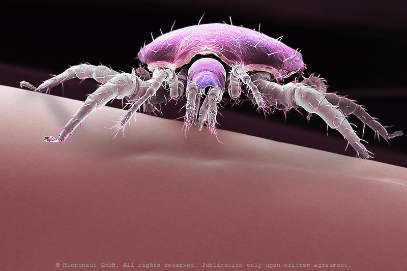
Watermite (Hydrachna sp.), Nr.1
Prostigmata: Hydrachnidae: Hydrachna sp. (a water mite predatory on the eggs of aquatic Heteroptera). With long swimming hairs on rear legs.
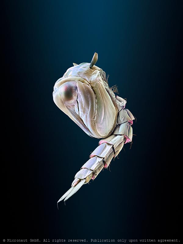
Divided Eye (Gyrinus substriatus)
Portrait of a Whirligig Beetle (Gyrinus substriatus). The most interesting insect compound eyes are those of whirligig beetles (Family Gyrinidae). You've probably seen groups of these lively little metallic beetles on ponds and slow streams. They have two sets of compound eyes. One pair of eyes is on top of the head to see what's going on above, i.e. to watch for predators. The other pair of eyes is located below the water surface to look out for smaller aquatic insects on which they prey on themselves. The antennae are also divided which is very unusual among beetles: the short and plump part is placed about at water level while the hairy lower part remains permanently submerged. The long bristles are used to detect vibrations of struggling insects that have fallen in and float with the current.
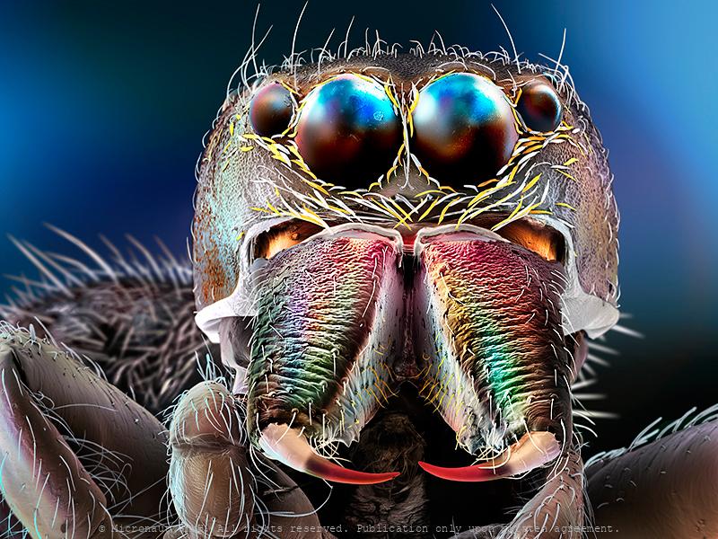
Jumping Spider
Often fuzzy and measuring less than a half inch in body length, Jumping spiders (Salticids) can run, climb, and - jump! Thereby the small predators can achieve stunning distances of more than 50 times its body size. Surprisingly, the extreme jumps are not the result of very strong or muscular legs. Instead, Jumping spiders quickly increase the blood pressure to their legs, which causes the legs to extend and propel the spider through the air. Prior to jumping, whatsoever, the spider glues silk threads to the surface beneath, to allow a quick and safe return to the perch, if required. Interestingly, Salticids rely on an excellent visual system. Like most other spiders, they have eight eyes on their face. An enormous pair in the center facing towards the front, and one adjacent pair of eyes to both sides give them an almost alien appearance. The remaining smaller eyes are located on the dorsal surface of the cephalothorax and allow almost 360° angle panoramic view. In general, animals have developed several different visual systems to accurately and reliably judge distance and depth. Humans, for example, have binocular stereovision. Because our eyes are spaced apart, they receive visual information from different angles, which our brains use to automatically triangulate distances. Other animals, such as insects, adjust the focal length of the lenses in their eyes, or move their heads side to side to create an effect called motion parallax — nearer objects will move across their field of vision more quickly than objects farther away. However, Jumping spiders have eyes that are too close together for binocular stereovision and don’t appear to use motion parallax while hunting. So how are these creatures able to perceive depth? In fact, these amazing creatures have developed a way of gauging distances that appears to be unique in the animal kingdom: rather than having a single layer of photoreceptor cells, as in our case, the spider's retina of the principal e
Turban Eye (Ephemeroptera)
Mayflies produce eggs that are laid on the surface of lakes or streams where they sink to the bottom. Naiads moult 20-30 times over a period of few months up to one year or even more. They live primarily on algae or diatoms, but some species are also predatory. The lifespan of adult mayflies is very short and varies between hours and 2-3 weeks. Since the primary function of the adult is reproduction, the mouthparts are non-functional and the digestive tract is filled with air. Adults have short flexible antennae, usually three ocelli and relatively large compound eyes. In most species, males additionally feature very large turban-shaped dorsal eyes, i.e. greatly enlarged compound eyes that are located on top of the head. The dorsal turban-eyes are especially designed to boost light sensitivity and allow vision at low light. At dawn, male mayflies fly close to the water surface in search of females, trying to detect females against the dim background of the sky. The turban-eyes are only capable of detecting ultra-violet (UV) light. In some species (e.g. European Mayfly) the turban-eye of the male is divided: The upper portion is for seeing movement, and the lower portion is specialized for seeing details. The females have smaller eyes.
Moth head
Understanding other animals’ sensory abilities allows us to put our capabilities and limitations into perspective and lets us appreciate what we have. It also lets us imagine what it would be like to perceive the world through another creature’s eyes and - inspires technological innovations (ie: when designing robots or night vision goggles). In general the insect compound eye is made up of many separate lenses, called ommatidia. Moths have huge eyes covering almost their entire head. The huge compound eye even lets them see 'in the back of their head' at the cost of a relatively low resolution, because it depends from the number of photoreceptors (i.e. number of lenses) that can be grouped on the relatively small head. Thereby insects perceive the environment as 'patchwork image', as if we would look through the grid of a cellar window. Nevertheless the small moth has a few tricks up their sleeves for a better vision in the dark: (1) Neural summation: moths' brains store up images over time, giving their photoreceptors more chances to see in dim light (our photoreceptors 'stream' images in a constant flow which means old images are almost immediately lost). (2) Ultraviolet photoreceptor type. Instead of red, moths have ultraviolet photoreceoptor types helping them to find flowers at night: in contrast to humans, moths rely on color vision even at very low light without having to switch to achromatic vision to differentiate colors at low intensities. It may give them a less accurate sense of color at daylight, but moths see color at much lower intensities than humans. (3) Antireflective coatings: the ommatidia of moths' are covered with micro-structures which increases light efficiency at night and helps to avoid predators at daylight (frogs, lizards, birds). Anti-reflective coatings have become a key research area in solar panel construction, because they increase the efficiency.
Rat flea (Xenopsylla cheopis)
In general, fleas belong to the insect order Siphonaptera. Their color varies between pale yellow and black and they are rather smalle, usually between 1-8mm long. The heavily sclerotized (armored) body is shiny. The rat flea's primary host is the rat (Rattus norvegicus), but the Oriental rat flea is best known as the vector for the bacteria Yersinia pestis from rats to humans, and for spreading the resulting plague (black death) across Asia and Europe in the middle ages. In the 14th century (between 1347 and 1352), the disease killed 25 million in Europe and between 75 to 100 million worldwide, within five years. Today, the plague is still not wiped out completely and the WHO registers annually between 1'000-3'000 cases worldwide. Like all fleas, Xenopsylla cheopis has mouthparts adapted to cutting through skin and sucking up blood that has pooled. In feeding, it secretes saliva into the wound to prevent the blood from coagulating. Along with the saliva, the flea secretes any bacteria it may have picked up by eating the blood of an infected individual into the host. When Yersinia pestis pathogens enter the gut of the flea, they multiply quickly, blocking food from entering the digestive system. This triggers the hungry flea to bite a new host, further spreading the bacteria. Today the Oriental rat flea can be found in temperate climates, but more often inhabits warm, tropical and subtropical regions, since it needs warm temperatures to pupate. It uses many different mammals as hosts, including rats and humans, and is known also to carry the murine typhus pathogen (Rickettsia typhi) and the mouse and rat tapeworms (Hymenolepis diminut and Hymenolepis nana), the latter can be transmitted when fleas are swallowed by pets or humans.
Catflea (Ctenocephalides felis), Nr.2
Frontansicht eines Katzenflohs. Parasiten sind normalerweise auf einen bestimmten Wirt spezialisiert. Katzenflöhe auf Katzen, nur ausnahmsweise werden auch Menschen gestochen (wenn keine Katze in der nähe ist). Portrait of a cat flea (magn. 158:1 if printed 12.5cm high). The cat flea's primary host is the domestic cat, but this is also the primary flea infesting dogs in most of the world. Cat fleas can transmit other parasites and infections to dogs and cats and also to humans. The most prominent of these are Bartonella, murine typhus, and apedermatitis. The tapeworm Dipylidium caninum can be transmitted when a flea is swallowed by pets or humans. In addition, cat fleas have been found to carry Borrelia burgdorferi, the etiologic agent of Lyme disease, but their ability to transmit the disease is unclear.
Cat Flea (Ctenocephalides felis), Nr.7
Catflea (Ctenocephalides felis), Nr.2
Frontansicht eines Katzenflohs. Parasiten sind normalerweise auf einen bestimmten Wirt spezialisiert. Katzenflöhe auf Katzen, nur ausnahmsweise werden auch Menschen gestochen (wenn keine Katze in der nähe ist). Portrait of a cat flea (magn. 158:1 if printed 12.5cm high). The cat flea's primary host is the domestic cat, but this is also the primary flea infesting dogs in most of the world. Cat fleas can transmit other parasites and infections to dogs and cats and also to humans. The most prominent of these are Bartonella, murine typhus, and apedermatitis. The tapeworm Dipylidium caninum can be transmitted when a flea is swallowed by pets or humans. In addition, cat fleas have been found to carry Borrelia burgdorferi, the etiologic agent of Lyme disease, but their ability to transmit the disease is unclear.
Uneven brothers (D. melanogaster), Nr.2
Coloured scanning electron micrograph (SEM) of two mutant Fruitfly (Drosophila melanogaster) heads. Wildtype (i.e. normal) Fruitflies have two compound eyes (red) - one on either side of the head. Small bristles between the single lenses of the eye make sure it cannot be covered with dust or dirt particles. Genetically manipulated Flies can either lack compound eyes completely (left) or have additional eyes on the antennae, legs and other body areas (right). Fruit flies are widely used in genetic experiments, particularly in mutation experiments, because they reproduce rapidly and their genetic systems are well understood. This image visualizes how easily the results of the genetic modification can be observed in the Fruitfly which is one of the main reasons why it is still the most frequently used model organism in genetics despite more than 100 years of experimental research. left: Sine oculis-1 (so1) variant which lacks the compound eyes completely. right: Eyeless variant with ectopically expressed compound eyes under dpp-promotor in all imaginal discs. Handkoloriertes Raster-Elektronen-Mikroskopiebild zweier Fruchtfliegen-Mutanten. Die Taufliege oder Fruchtfliege (Drosophila melanogaster) ist das klassische Untersuchungsobjekt in der Genetik und Entwicklungsbiologie. Wildtypen, d.h. genetisch unveränderte Fruchtfliegen, besitzen zwei grosse rote Komplexaugen - je eines auf jeder Seite des Kopfes. Die Komplexaugen werden durch Borsten zwischen den winzigen Einzelaugen vor Verschmutzung und Staub geschützt. Die Entwicklung des Auges wird während der Embryonalentwicklung Larve durch bestimmte Gene gesteuert. Durch Veränderungen (Mutation) von Genen lassen sich z.B. Position oder Morphologie der Komplexaugen gezielte beeinflussen. Im Vergleich zu einem Säugetier ist die Fruchtfliege genetisch viel einfacher aufgebaut und weist lediglich vier Paar Chromosomen auf. Diese Tatsache hat, neben der raschen Generationsfolge und einfachen Zucht, wesentlich dazu
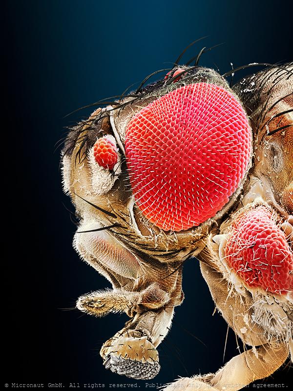
Uneven brothers (D. melanogaster), Nr.1
Coloured scanning electron micrograph (SEM) of two mutant Fruitfly (Drosophila melanogaster) heads. Wildtype (i.e. normal) Fruitflies have two compound eyes (red) - one on either side of the head. Small bristles between the single lenses of the eye make sure it cannot be covered with dust or dirt particles. Genetically manipulated Flies can either lack compound eyes completely (left) or have additional eyes on the antennae, legs and other body areas (right). Fruit flies are widely used in genetic experiments, particularly in mutation experiments, because they reproduce rapidly and their genetic systems are well understood. This image visualizes how easily the results of the genetic modification can be observed in the Fruitfly which is one of the main reasons why it is still the most frequently used model organism in genetics despite more than 100 years of experimental research. left: Sine oculis-1 (so1) variant which lacks the compound eyes completely. right: Eyeless variant with ectopically expressed compound eyes under dpp-promotor in all imaginal discs. Handkoloriertes Raster-Elektronen-Mikroskopiebild zweier Fruchtfliegen-Mutanten. Die Taufliege oder Fruchtfliege (Drosophila melanogaster) ist das klassische Untersuchungsobjekt in der Genetik und Entwicklungsbiologie. Wildtypen, d.h. genetisch unveränderte Fruchtfliegen, besitzen zwei grosse rote Komplexaugen - je eines auf jeder Seite des Kopfes. Die Komplexaugen werden durch Borsten zwischen den winzigen Einzelaugen vor Verschmutzung und Staub geschützt. Die Entwicklung des Auges wird während der Embryonalentwicklung Larve durch bestimmte Gene gesteuert. Durch Veränderungen (Mutation) von Genen lassen sich z.B. Position oder Morphologie der Komplexaugen gezielte beeinflussen. Im Vergleich zu einem Säugetier ist die Fruchtfliege genetisch viel einfacher aufgebaut und weist lediglich vier Paar Chromosomen auf. Diese Tatsache hat, neben der raschen Generationsfolge und einfachen Zucht, wesentlich dazu
Pseudoscorpion (Chelifer cancroides), Nr.2
Pseudoscorpions are small arachnids with a flat, segmented, pear-shaped body. Despite being harmless and inoffensive creatures, this individual was able to move very quickly to escape. Pseudoscorpions got their name from a pair of huge pincers (pedipalps), resembling those of scorpions. But unlike true Scorpions they have a rounded abdomen that does not extend into a segmented tail with a stinger. The small creatures usually range from 2 to 8 millimetres (0.08 to 0.31 in) in length, with yellowish to red or dark brown colors. Pseudoscorpions are generally beneficial to humans and prey on clothes moth larvae, carpet beetle larvae, booklice, ants, mites, and small flies. Chelifer cancroides is the most commonly found species in homes, where they are often observed in rooms with dusty books. A venom gland and duct are located in the mobile finger of the pincers. The venom is used to capture and immobilize prey. During digestion, a mildly corrosive fluid is poured over the prey to liquify the remains externally before they are ingested. They enter homes by "riding along" attached to insects (known as phoresy). The jaws (chelicera; have a look at the beautiful structure in the center) are used to spin silk which is used to make disk-shaped cocoons for mating, molting, or waiting out cold weather. A comb-like structure (serrula externa), which can be seen on the left side of the chelicera in a close-up picture, is used for grooming. In the center of the chelicera is a membrane called serrula interna. It helps to prevent food and fluids from escaping. Also, two pairs of sensory setae can be found on the chelicera and additional structures which resemble plant pores are located on the cephalothorax (above the eyes in the overview picture). Scientists believe that the latter detect bending and stress on the cuticule and call them lyrifissures. Pseudoscorpions breathe through spiracles on their abdomen (not shown), a trait they share with the insects. Most species have o
Pseudoscorpion (Chelifer cancroides), Nr.1
Pseudoscorpions are small arachnids with a flat, segmented, pear-shaped body. Despite being harmless and inoffensive creatures, this individual was able to move very quickly to escape. Pseudoscorpions got their name from a pair of huge pincers (pedipalps), resembling those of scorpions. But unlike true Scorpions they have a rounded abdomen that does not extend into a segmented tail with a stinger. The small creatures usually range from 2 to 8 millimetres (0.08 to 0.31 in) in length, with yellowish to red or dark brown colors. Pseudoscorpions are generally beneficial to humans and prey on clothes moth larvae, carpet beetle larvae, booklice, ants, mites, and small flies. Chelifer cancroides is the most commonly found species in homes, where they are often observed in rooms with dusty books. A venom gland and duct are located in the mobile finger of the pincers. The venom is used to capture and immobilize prey. During digestion, a mildly corrosive fluid is poured over the prey to liquify the remains externally before they are ingested. They enter homes by "riding along" attached to insects (known as phoresy). The jaws (chelicera; have a look at the beautiful structure in the center) are used to spin silk which is used to make disk-shaped cocoons for mating, molting, or waiting out cold weather. A comb-like structure (serrula externa), which can be seen on the left side of the chelicera in a close-up picture, is used for grooming. In the center of the chelicera is a membrane called serrula interna. It helps to prevent food and fluids from escaping. Also, two pairs of sensory setae can be found on the chelicera and additional structures which resemble plant pores are located on the cephalothorax (above the eyes in the overview picture). Scientists believe that the latter detect bending and stress on the cuticule and call them lyrifissures. Pseudoscorpions breathe through spiracles on their abdomen (not shown), a trait they share with the insects. Most species have o
The First Dsease of Humans (Scabies, 1687)
This is a hand colored scanning electron micrograph (SEM) of Sarcoptes scabiei, the itch mite, which is a parasitic arthropod burrowing into human skin, thereby causing scabies. The discovery of the itch mite in 1687 marked scabies as the first disease of humans with a known cause. Other mammals can also become infected, including dogs, cats, ungulates, wild boars, bovids, wombats, koalas, and great apes. About 2% of the British population is thought to be infected with these mites, which take about 25 minutes to an hour to burrow into the skin. The burrowing is carried out using the mouth parts and special cutting surfaces on the front legs. A burrowing mite anchors with suckers on its feet, furrows and spines, located on its body surface. Eggs are laid in small numbers as the mite burrows. As these hatch, six-legged larvae climb out on to the skin and search for hair follicles, where they feed and develop. In the hair follicles, the larvae show the first nymphal stages, with eight legs.
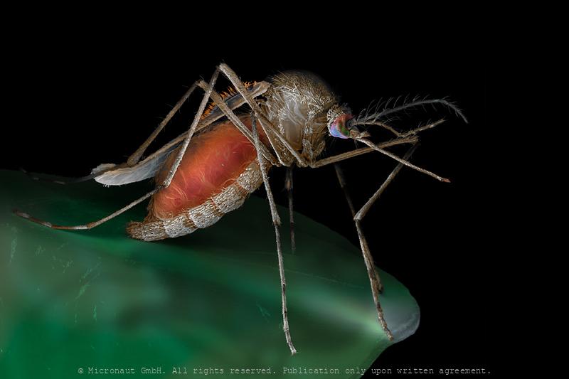
Mosquito after a meal (Culex pipiens)
Adult Mosquito after a meal (Culex pipiens).
Born to Buzz - The Mosquite Wing (Anopheles gambiae)
Detail of a Mosquito wing. We are all familiar with the annoying high frequency sound of Mosquitoes. They have evolved unique wing motions and distinct wing shapes to maximize the sound power during flight: - their wing flapping frequency is remarkably high (>700 Hz) which generates a high frequency wing buzz - in addition, the wing stroke angle of the mosquito is much smaller than for other insects. The stroke angle of the mosquito (Culex, male) is only about 40 degrees, whereas it is about 150 degrees for the fruit fly. For a given mechanical energy input, mosquitoes maximize their sound power through these conditions. Acoustic communication is very important in the lifecycle of mosquitoes - which are important vectors for some of the most devastating diseases that afflict mankind.
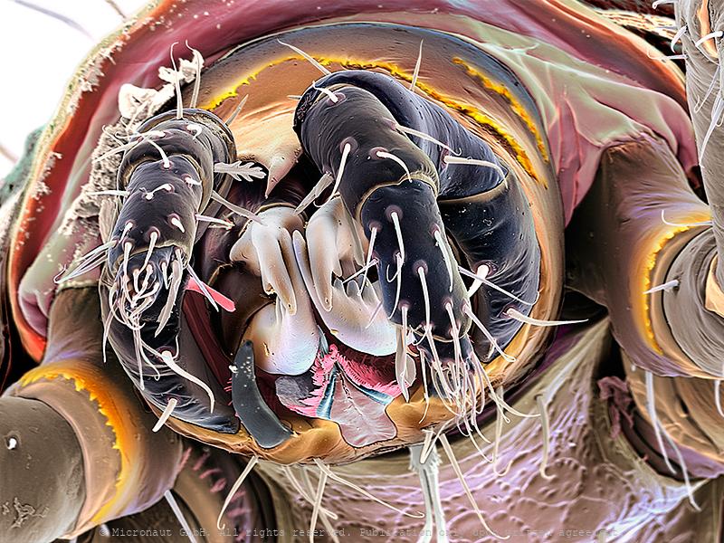
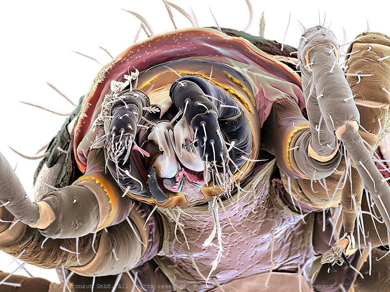
Head louse (Pediculus humanus capitis), Nr.1
Head louse (Pediculus humanus capitis) parasitize humans for a long time. Genetic analysis suggest that the ancestors of the lice may have originated about 107’000 years ago, with the ancestor of all human lice emerging about 770’000 years ago. The Head louse is an obligate ectoparasite of humans and spends the entire life cycle on the human scalp in contrast to other human parasites, e.g. fleas. Lice are wingless insects and they feed exclusively on blood. Unlike Body lice, Head lice are not the vectors of any known diseases. Except for rare secondary infections that result from scratching at bites, head lice are harmless, and they have been regarded by some as essentially a cosmetic rather than a medical problem. It has even been suggested that head lice infections might be beneficial in helping to foster a natural immune response against lice which helps humans in defense against the far more dangerous body louse, which is capable of transmission of dangerous diseases.
Head louse (Pediculus humanus capitis), Nr.2
Head louse (Pediculus humanus capitis) parasitize humans for a long time. Genetic analysis suggest that the ancestors of the lice may have originated about 107’000 years ago, with the ancestor of all human lice emerging about 770’000 years ago. The Head louse is an obligate ectoparasite of humans and spends the entire life cycle on the human scalp in contrast to other human parasites, e.g. fleas. Lice are wingless insects and they feed exclusively on blood. Unlike Body lice, Head lice are not the vectors of any known diseases. Except for rare secondary infections that result from scratching at bites, head lice are harmless, and they have been regarded by some as essentially a cosmetic rather than a medical problem. It has even been suggested that head lice infections might be beneficial in helping to foster a natural immune response against lice which helps humans in defense against the far more dangerous body louse, which is capable of transmission of dangerous diseases.
Head louse (Pediculus humanus capitis), Nr.3
Head louse (Pediculus humanus capitis) parasitize humans for a long time. Genetic analysis suggest that the ancestors of the lice may have originated about 107’000 years ago, with the ancestor of all human lice emerging about 770’000 years ago. The Head louse is an obligate ectoparasite of humans and spends the entire life cycle on the human scalp in contrast to other human parasites, e.g. fleas. Lice are wingless insects and they feed exclusively on blood. Unlike Body lice, Head lice are not the vectors of any known diseases. Except for rare secondary infections that result from scratching at bites, head lice are harmless, and they have been regarded by some as essentially a cosmetic rather than a medical problem. It has even been suggested that head lice infections might be beneficial in helping to foster a natural immune response against lice which helps humans in defense against the far more dangerous body louse, which is capable of transmission of dangerous diseases.
©-Micronaut-Headlice-PTUmini
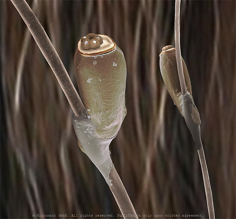
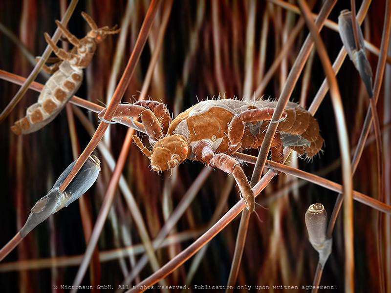
Body lice eggs
Lice egg and
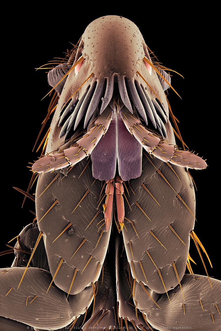
Predatory Mite (Gamasellus sp.), Nr.2
Colored Scanning Electron Micrograph of a predatory mite (Gamasellus sp.). You can be grateful this animal measures only app. 0.18mm in diameter, because it has a horrible bite: adult Gamasellus eat Springtails (Collembola sp.) of the same size! Compared to juveniles, adults focus on larger prey and eat more often (juvenile: 1 Springtail every 12 days, adult: 1 Springtail every 3 days). Gamasellus is an agile predator and attacks quickly through looping his elongated forelegs over the prey. After this, the chelicerae are moved forward to "kiss" the prey, and inject digestion fluid through a pinhole at the tip of the upper left and right chelicerae. The poisonous fluid tranquilizes and eventually kills and digests the prey. Such a procedure is called pre-oral digestion and it is common in the class of Arachnida, especially in spiders. A multitude of mechanosensory setae ("hairs") are located on the forelegs and pedipalps. They serve the blind predator to monitor the exact position of its prey and make sure it is held in position until the deadly cocktail is effectively working.
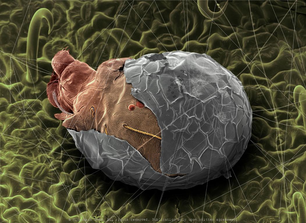
Hatching spider mite
Hatching spider mite
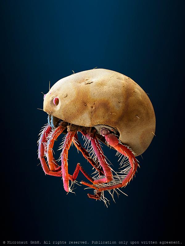
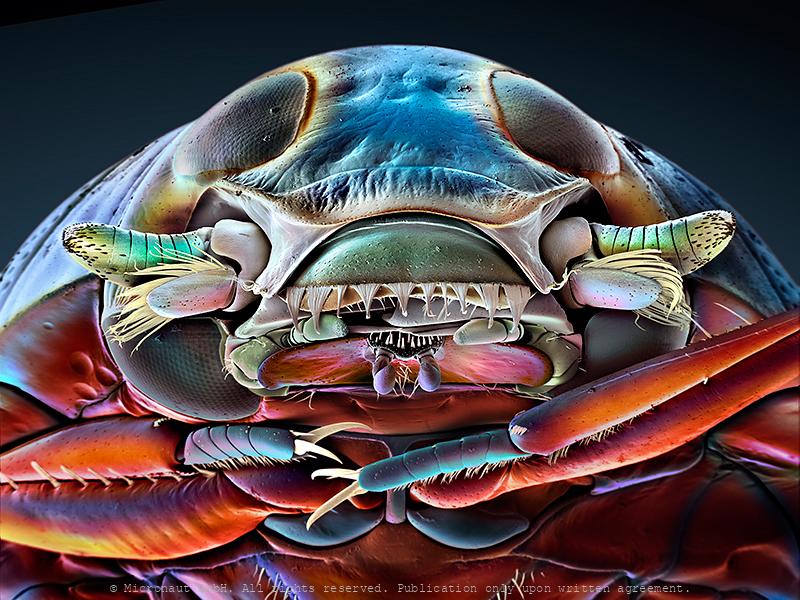
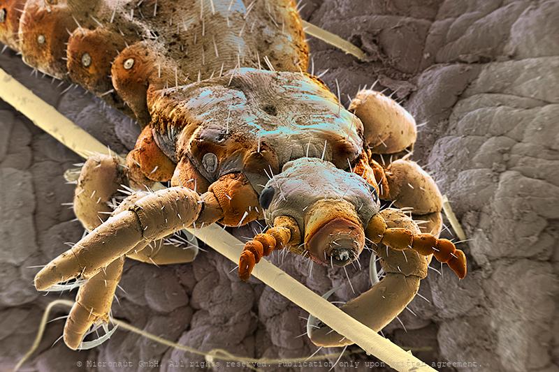
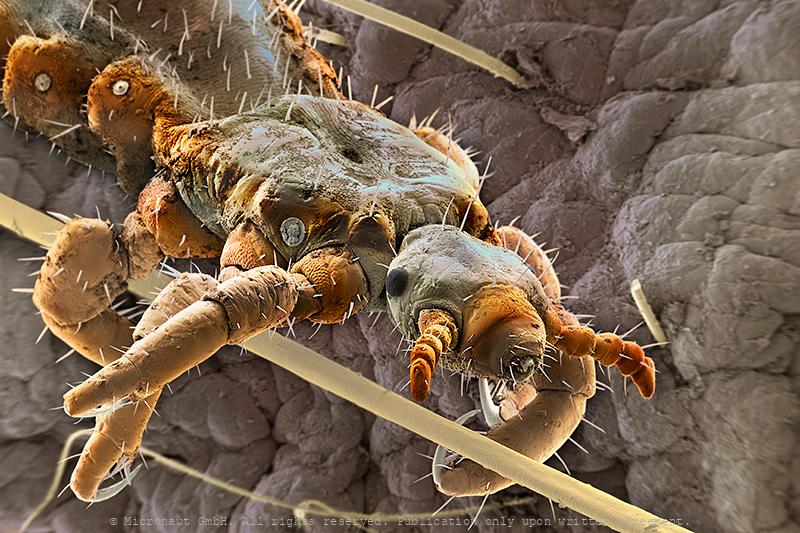
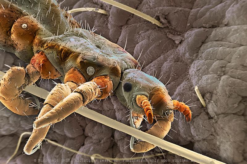
Parasitic mite (unknown species)
Catflea (Ctenocephalides felis), Nr.1
Flea portrait
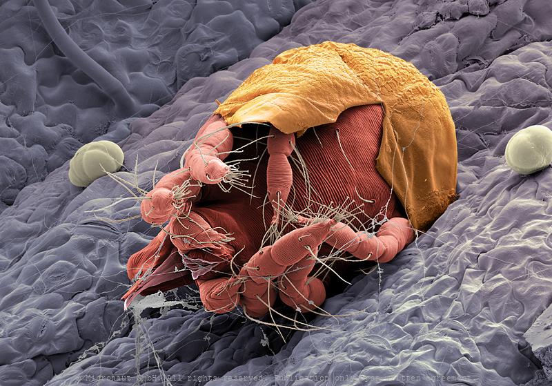
Hatching spider mite
Hatching spider mite
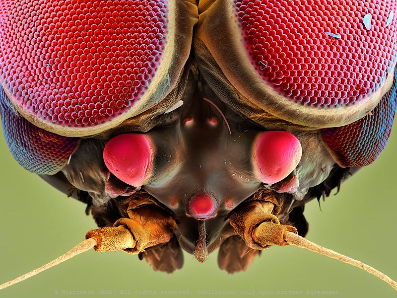
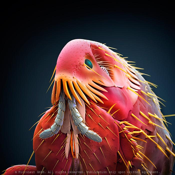
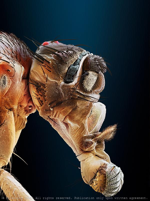
fly head
Fly head (Drosophila melanogaster)